



Digital Modeling Hub
Filters
-
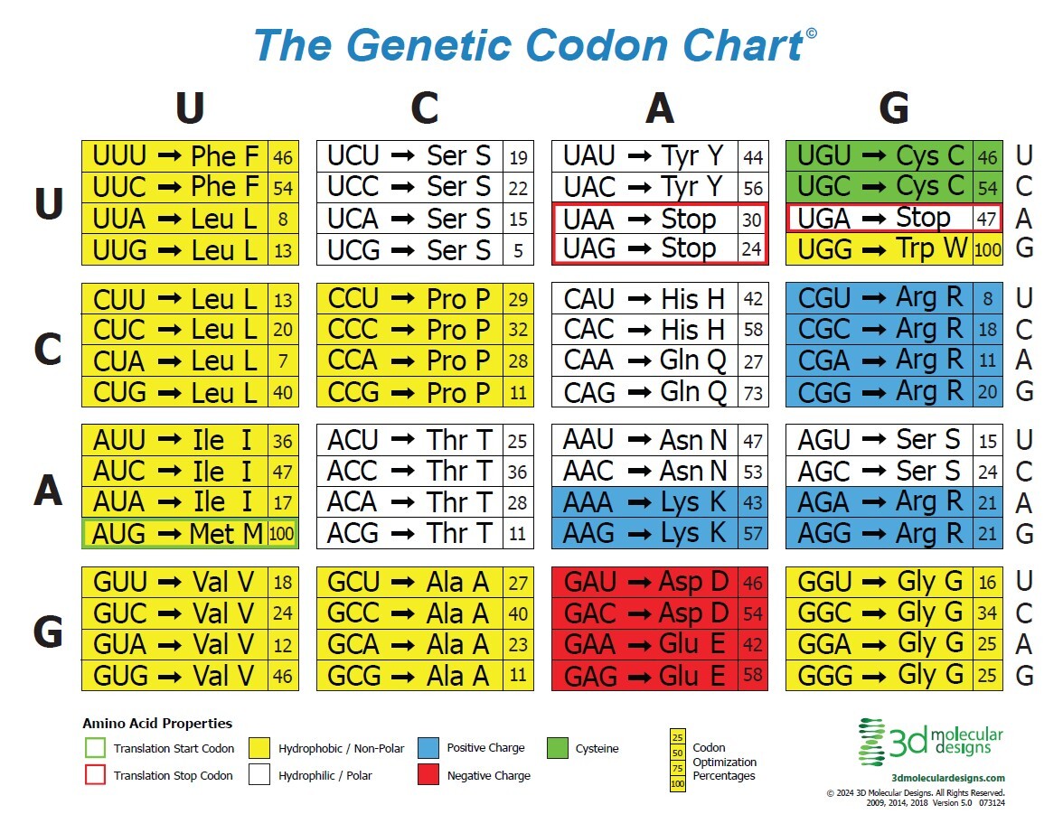
Genetic Codon Chart Activities & Resource Guide


![]() Associated Product:
Associated Product:
Flow of Genetic Information Kit©Connect the big picture of the central dogma to the codon-by-codon decisions made during translation! The Flow of Genetic Information Kit© already guides students through replication, transcription, and translation, including decoding mRNA into protein on the ribosome placemat. This insert adds a focused pause point where students use the Genetic Codon Chart (Circle or Table) to strengthen their understanding of how the sequence of mRNA nucleotides determines the sequence of amino acids in proteins.
-
Organ Systems at Work Slide Deck
![]() Associated Product:
Associated Product:
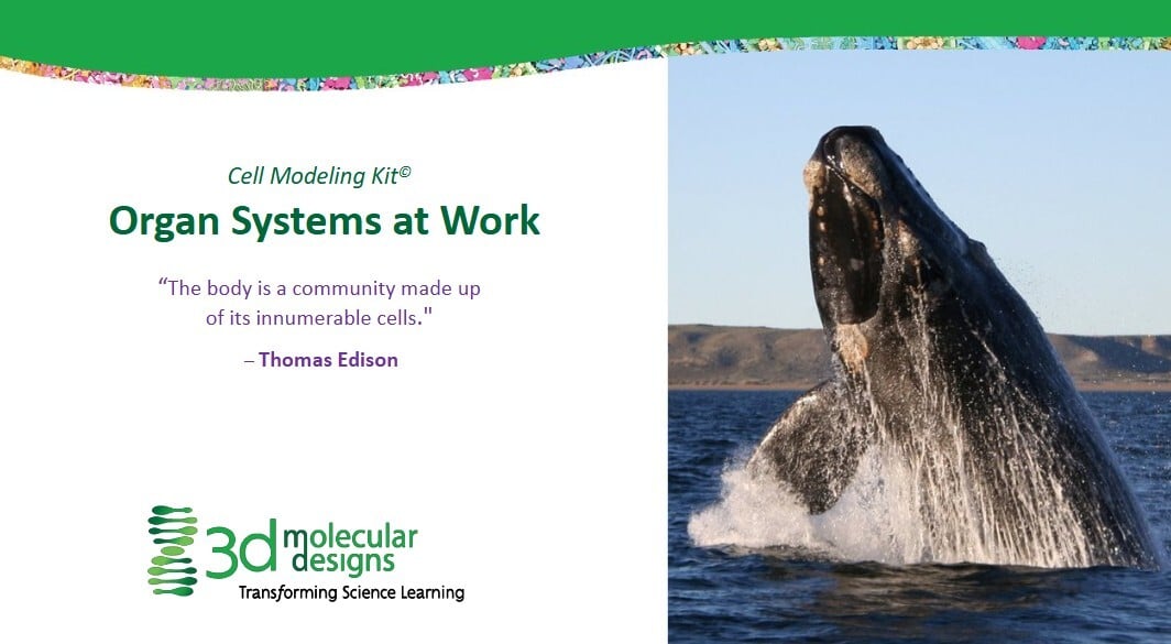
Cell Modeling Kit©Organ Systems at Work is a 60–75 minute, hands-on modeling lesson that helps students visualize how cells build up into tissues, organs, and organ systems in multicellular organisms. Students use the Cell Modeling Kit© in groups of 4–6 to first complete a free-build of a cell, activating prior knowledge about cell structures and functions and then sharing and comparing models through a gallery walk to surface similarities, differences, and common misconceptions. Guided notebook drawing, labeling, and discussion push students to connect cell structure to function and to consider how many diverse cell types are needed for a large organism such as a blue whale to survive.
From there, students move explicitly into levels of organization: they learn that cells with similar structure and function form the four major animal tissue types (epithelial, connective, muscle, and nervous), and that these tissues combine to form organs, which in turn coordinate in organ systems to keep an organism healthy. Across three sequenced activities—reviewing and modeling cell types, comparing cells and tissues, and reasoning about how many different cells must work together as a living thing—students develop and revise models, analyze patterns, and construct explanations about structure–function relationships from the cellular through the organismal level. The slide deck is written for middle school life science and tightly aligns with NGSS MS-LS1-3 and related SEPs (Developing and Using Models, Constructing Explanations) and CCCs (Structure and Function, Patterns, Systems), making it a strong fit for units on cells, body systems, or levels of organization in grades 6–8, and adaptable as an anchoring or reinforcement activity in high school biology or anatomy & physiology. The activity's emphasis on three-dimensional manipulation of molecular/cellular models makes it particularly effective for visual and kinesthetic learners who benefit from handling physical representations of microscopic structures.
-
Building the Perfect Cell: How Can a One-celled Euglena Live Slide Deck
![]() Associated Product:
Associated Product:
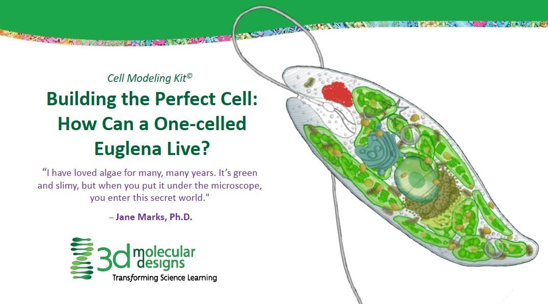
Cell Modeling Kit©Your students will use the Cell Modeling Kit© to explore the structure and function of cellular organelles in prokaryotic and eukaryotic cells. The 90-minute lesson engages students with a constructivist approach: they begin by building their own "perfect cell" without instruction, then engage in gallery walks to compare designs, and finally learn about specific organelles through guided instruction while reconstructing models. By starting with a free-build activity, students develop their own hypotheses about what cells need to survive, making later instruction more meaningful. The activity is scaffolded using the 5 E's learning model (Engage, Explore, Explain, Elaborate, Evaluate/Extend) and includes prompting icons that cue students to different types of thinking and interaction (turn-and-talk discussions, quiet reflection, physical and digital modeling).
This activity is designed for middle school life science (targeting 8th grade standards), with prerequisite concepts from 6th and 7th grade. It directly addresses NGSS standard MS-LS1-2 ("Develop and use a model to describe the function of a cell as a system whose properties and systems provide it with food, water, and energy") and aligns with the science and engineering practices of developing and using models, asking questions, and constructing explanations. The cross-cutting concept of "Structure and Function" is central throughout. The activity could also be adapted for high school biology courses and introductory college bioscience courses that cover cell biology, as the depth of detail about organelle functions and the comparison of cell types provides content suitable for more advanced students.
-
Antibody Production and Cellular Processes Slide Deck
![]() Associated Product:
Associated Product:
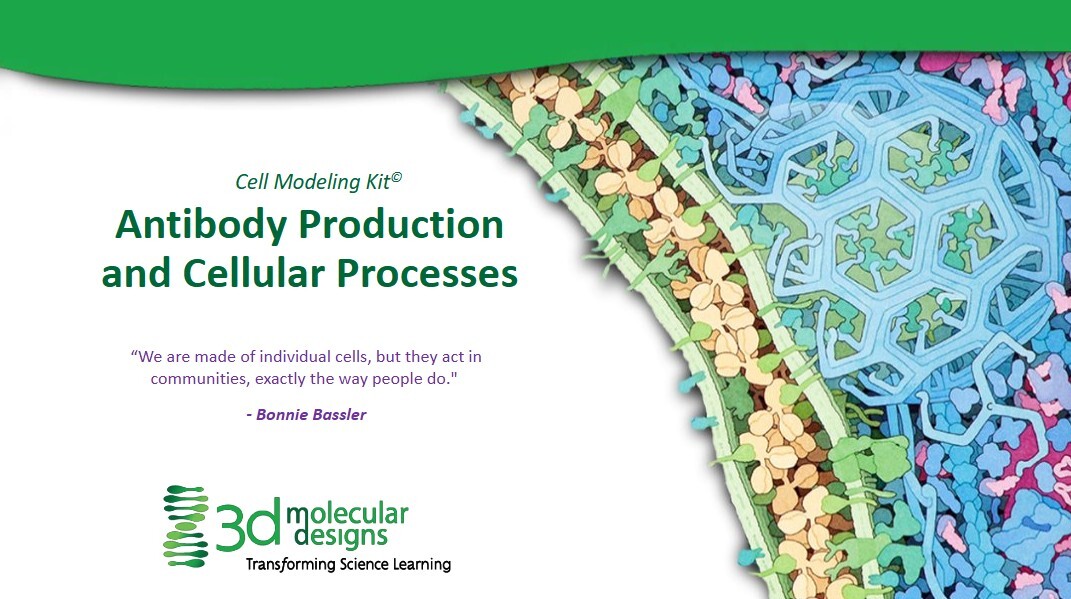
Cell Modeling Kit©Students develop a 3D B-cell model to follow an antibody’s “life story” from gene to secretion, making the central dogma and organelle functions concrete and memorable. Working in groups of four with the Cell Modeling Kit, they build a cell membrane and nucleus, then model transcription, translation, protein maturation and trafficking, and finally vesicle-mediated secretion of antibodies. At each step, students identify which organelles are involved, what is produced, and where it goes next, recording their thinking in lab notebooks and using prompts that surface and correct common misconceptions (for example, that DNA leaves the nucleus or that only one organelle is responsible for protein synthesis).
The slide deck includes detailed teacher notes with suggested prompts, checks for understanding, and timing, plus icons that signal student tasks such as discussion, notebook writing, and quiet thinking. Optional extensions invite students to use the same modeling approach to explore additional processes such as glucose update and signal transduction, cell replication and division, virus infection cycle, neurotransmission and action potential, macrophage or neutrophil endocytosis, and protein degradation and recycling, making the resource flexible for a single class period or a short multi-day sequence.
Best fit: 9–12 Biology, Honors Biology, and AP Biology classes addressing cell structure and function, protein synthesis, or the immune system. This activity directly supports NGSS HS-LS1-2 and HS-LS1-6 with a strong focus on developing and using models and on structure–function relationships in cells.
-
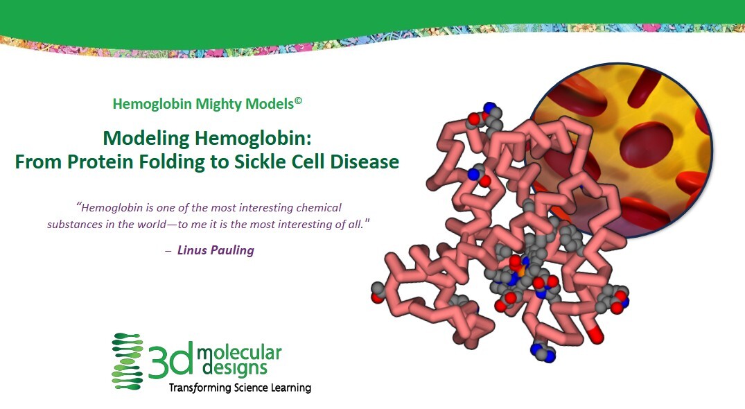
Modeling Hemoglobin: From Protein Folding to Sickle Cell Disease Slide Deck
![]() Associated Product:
Associated Product:
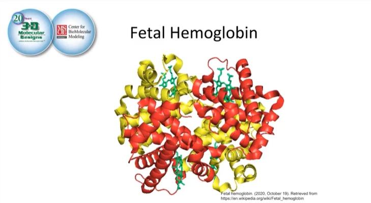
Hemoglobin Mighty Model©Students use 3D alpha- and beta-globin models to investigate all four levels of protein structure while connecting molecular details to a real patient story and the genetics of sickle cell disease. Anchored in the case of Victoria Gray, students begin at the organism and organ-system level (symptoms, circulation, red blood cell shape), then zoom in to the cellular and molecular scales to explore how hemoglobin’s structure enables oxygen transport—and how a single amino acid substitution (glutamic acid to valine at position six in beta-globin) can distort red blood cells and impair blood flow. Along the way, they trace the globin backbone from N- to C-terminus, link “bends” in the ribbon to individual amino acids, examine selected sidechains, and classify them as hydrophilic or hydrophobic to explain how polarity, charge, and the hydrophobic effect drive folding into tertiary and quaternary structures and position heme groups for oxygen binding.
Designed primarily for grades 9–12 Biology, Honors Biology, and AP Biology, this activity fits comfortably into one 60–75 minute class period for the core investigation, with an additional 45–60 minutes if you choose to use all extensions and assessments. It pairs especially well with 3DMD’s Amino Acid Starter Kit and Protein Student Modeling resources, giving students a concrete bridge from amino acid properties to full protein models. The slide deck includes student-facing prompts and rich teacher notes, with built-in routines such as Notice–Wonder charts, quiet thinking time, turn-and-talk discussion, digital and physical modeling, and frequent formative checks to surface misconceptions about protein structure, red blood cell function, and genetic disease.
For advanced high school or introductory college courses, optional extensions connect protein structure to gene structure and biotechnology. Students can map the 146–amino-acid beta-globin chain back to its gene with exons and introns, examine carbon monoxide as a competitive inhibitor at the heme site, and discuss how CRISPR-based genome editing has been used clinically to treat sickle cell disease, including in Victoria Gray’s case. These extensions support NGSS performance expectations HS-LS1-1 and HS-LS1-2 (structure and function of proteins), HS-LS3-1 and HS-LS3-2 (inheritance and variation of traits, including the Glu→Val mutation), and highlight crosscutting concepts of Structure and Function, Cause and Effect, and Systems and System Models, making the lesson a strong fit for units on molecular genetics, heredity, human physiology, and biotechnology.
-
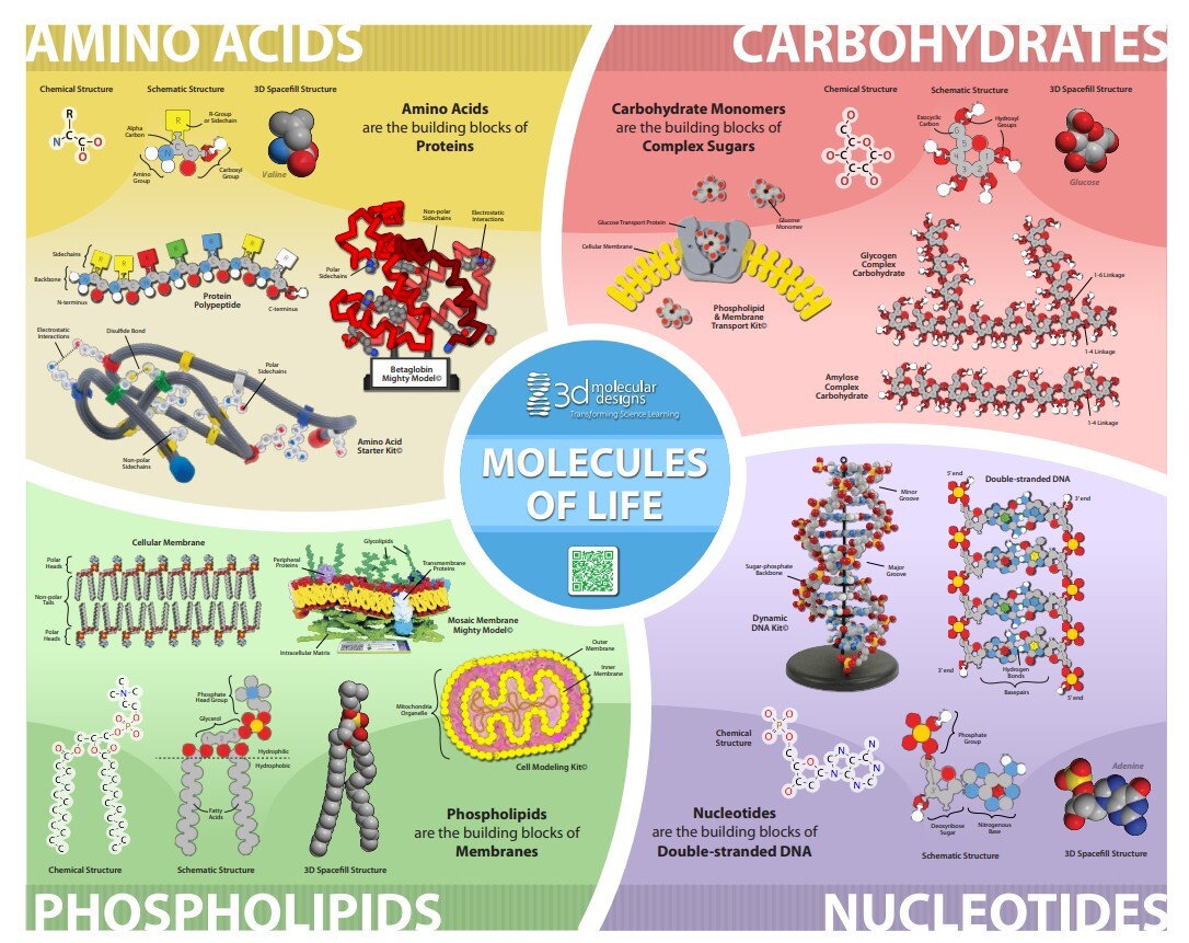
Molecules of Life Modeling Kit© Poster Teacher Resources
-
Building the Perfect Cell: Modeling the Characteristics of Life Activity Slide Deck
![]() Associated Product:
Associated Product:
Cell Modeling Kit©Your students will learn the characteristics and requirements of living cells through this hands-on modeling activity. Using a Cell Modeling Kit, student groups collaborate to construct models representing an ideal cell, ensuring their designs incorporate the critical criteria for life: metabolism, growth and development, response to stimuli, reproduction, adaptation, homeostasis, and protection. Throughout the lesson, students brainstorm the differences between living and nonliving things, create and refine their cell models, and develop a rubric to assess whether their models meet the requirements for life. The activity encourages iterative thinking by guiding students to modify their models in response to changing environmental scenarios, fostering a deeper understanding of cellular functions and scientific reasoning.
This lesson is aligned with Next Generation Science Standards (NGSS): MS-LS1-1 (Structure and Function), as students use models to explain how living things are made of cells, analyze evidence, and evaluate the necessary characteristics for life. The activity is well-suited for 6th-grade life science courses, but can be adapted for high school and introductory college biology classes focusing on cell biology and the nature of life. Beyond fostering teamwork and creativity, this engaging activity helps educators provide students with foundational knowledge of cells, supports inquiry-based learning, and allows for assessment through observation, discussion, and student-developed rubrics.
-
The Great Cell Build-Off: Unlocking Cell Specialization Activity Slide Deck
![]() Associated Product:
Associated Product:
Cell Modeling Kit©During the two-day sequence, students examine cell organelles, evaluate cellular differentiation, and connect these ideas to real-world examples like specialized animal and plant cells. Each step encourages observation, creative modeling, and group discussion—students document their work, compare findings, and reflect on how cell structures relate to their environment and function.
Using the Cell Modeling Kit©, students work in small groups to build models of prokaryotic cells, study their parts and functions, and compare them to mitochondria and chloroplasts. The lesson advances by guiding students through the endosymbiotic theory, leading them to create detailed models of eukaryotic cells and culminating in a focused discussion on how cells with specialized structures perform unique roles within multicellular organisms. This activity seamlessly aligns with the Next Generation Science Standards (NGSS), specifically HS-LS1-1 (Structure and Function), and emphasizes science practices such as modeling, evidence-based explanation, and recognizing patterns. Its flexibility also makes it adaptable for advanced middle school life science or introductory college biology courses.
-
Modeling the Molecules of Life Activity Slide Deck
![]() Associated Product:
Associated Product:
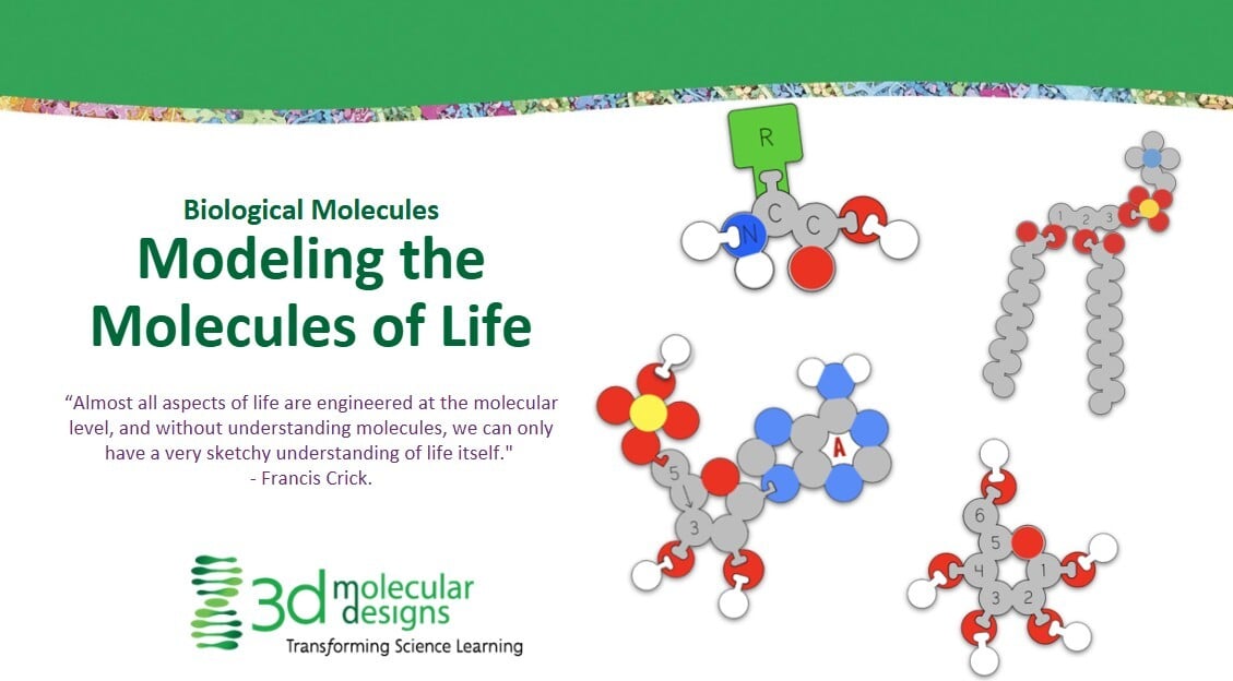
Molecules of Life Modeling Kit©Your students will use tactile modeling kits to build and analyze macromolecules, forming connections to real-life cell processes and health contexts. Students will explore the structure and function of biological macromolecules—proteins, lipids, carbohydrates, and nucleic acids. Students use physical models to demonstrate biochemical processes such as dehydration synthesis and hydrolysis, construct polypeptides, phospholipids, disaccharides, and nucleotide chains, and compare healthy and unhealthy cells by observing how biomolecular changes impact function. The approach emphasizes scientific practices from the NGSS, like developing and using models and constructing explanations, with targeted class discussions, drawing exercises, and written reflections to deepen understanding.
Recommended for grades 9-12 biology or chemistry courses, this lesson is easily adaptable for introductory college bioscience settings. It can be scaffolded for advanced middle school students with foundational knowledge of atoms, molecules, and covalent bonding. The activity is tightly NGSS-aligned, explicitly supporting HS-LS1.A (Structure and Function), HS-LS1-2 (Modeling and Explanation), and crosscutting concepts such as scale, proportion, and quantity. Educators can extend the lesson into topics like membrane transport, polysaccharide structure, DNA/RNA comparisons, and cancer biology, providing flexible entry points and extension opportunities for diverse science classrooms.
-
Molecules of Life Modeling Kit© - 4 Activity Slide Decks
![]() Associated Product:
Associated Product:
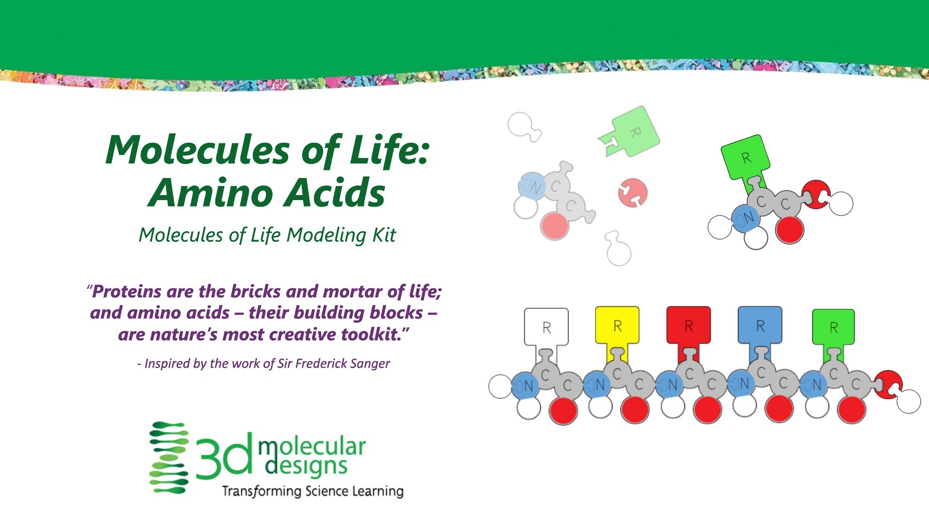
Molecules of Life Modeling Kit©Your students will investigate the four major types of biological macromolecules through hands-on modeling: amino acids (proteins), carbohydrates, nucleotides (DNA/RNA), and phospholipids (membranes) – in four activities. Using the Molecules of Life Modeling Kit, the lessons are structured to help students visually and physically build each type of molecular monomer and then assemble them into their respective polymers or supramolecular structures. For each molecule type, students identify the core chemical structures and variable side groups, model key biochemical processes (such as dehydration synthesis for polymer formation), and connect these molecular features to biological function.
Throughout the activities, your students engage in inquiry by observing and comparing models, noting similarities and differences between molecular structures, discussing their understanding, and sketching diagrams with labeled parts. The activities also prompt students to consider cross-cutting concepts such as the relationship between structure and function, the significance of repeating structural patterns, and the role of enzymes in biological synthesis. For amino acids, carbohydrates, and nucleotides, students model how covalent bonds link monomers into polymers, whereas for phospholipids, they explore how amphipathic properties lead to membrane formation without covalent polymerization. The slide decks emphasize active participation, group discussion, and reflection in lab notebooks to reinforce connections between molecular structure and its roles in living organisms.
-
Alpha Helix & Beta Sheet Structure Set© Stabilizing Structures Activity Slide Deck
![]() Associated Product:
Associated Product:
Alpha Helix & Beta Sheet Structure Sets©This activity is designed for high school biology classes (grades 9–12) and introduces students to the foundational concepts of protein structure, focusing on the secondary structures—alpha helices and beta sheets. Through hands-on exploration with physical models, students discover how proteins are composed of amino acids linked into chains, which then fold into unique shapes – alpha helices and beta sheets - stabilized by hydrogen bonds. The activity emphasizes the importance of these secondary structures as rigid scaffolds that support the overall 3D shape and function of proteins.
The detailed slide deck includes clear differentiation between student and teacher content, embedded prompts for various activity types (such as physical modeling and discussion), and teacher notes to guide facilitation. The activity requires no prerequisite knowledge, takes approximately 20 minutes, and aligns with Next Generation Science Standards (NGSS), specifically targeting modeling, explanation, and the relationship between structure and function in biological systems. For a deeper dive, an optional extension with the Amino Acid Starter Kit allows students to further investigate primary and tertiary protein structures by constructing and folding their own protein models.
-
Alpha Helix & Beta Sheet Structure Set© Protein Tertiary Structure Activity Slide Deck
![]() Associated Product:
Associated Product:
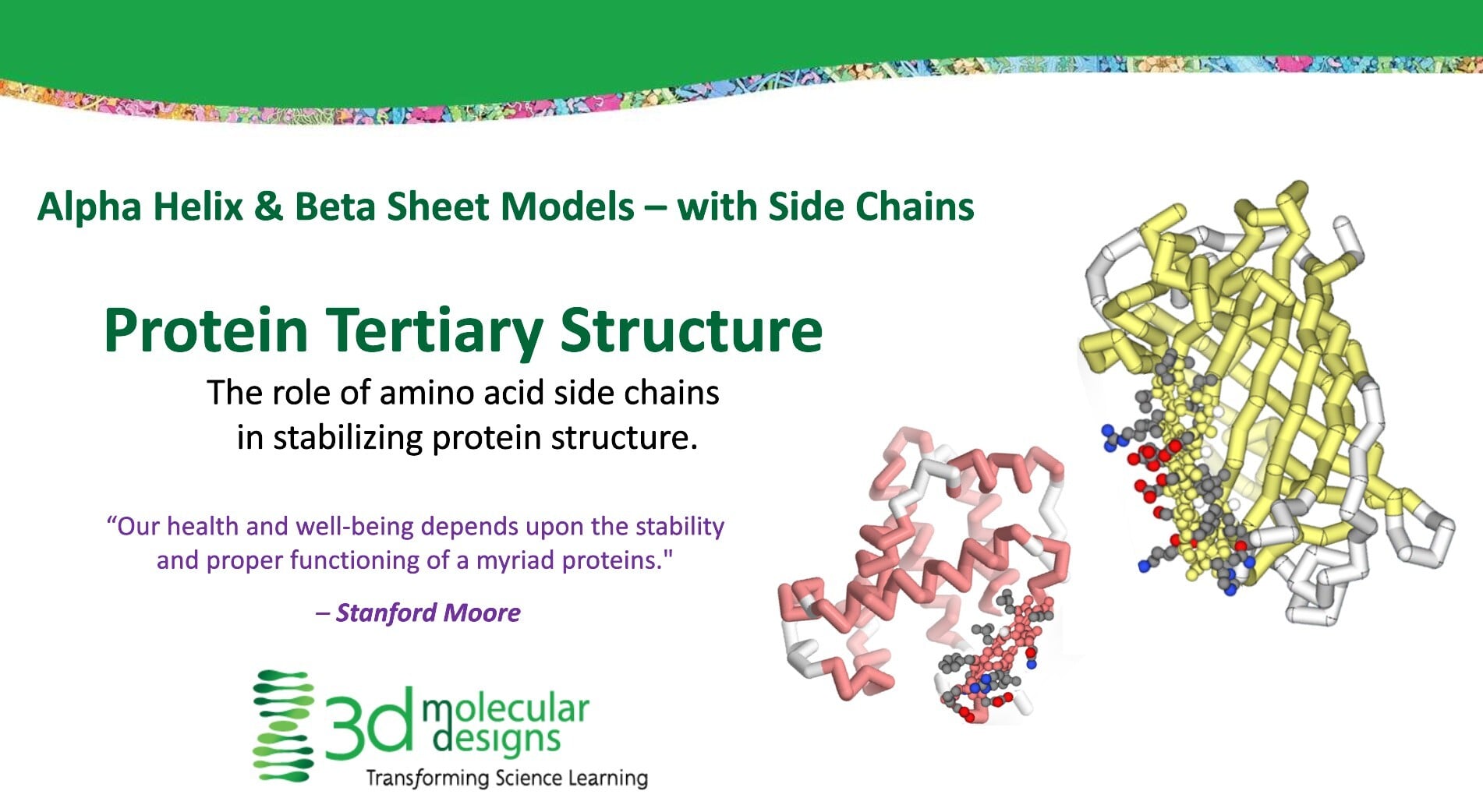
Alpha Helix & Beta Sheet Structure Sets©This activity introduces students to protein structure by exploring the role of side chains in alpha helices and beta sheets—in determining the tertiary structure of proteins. Through hands-on modeling, students learn that each protein folds into a unique three-dimensional shape, determined by the sequence and properties of its amino acids. The activity focuses on where and how amino acid sidechains (R-groups) attach to the protein backbone and how their chemical properties—hydrophobic or polar—influence protein folding and stability.
Students use physical models to compare simple amino acids to more complex protein structures, first observing models that display only the backbone and then examining models showing side chains. They identify which sidechains are hydrophobic or polar using a side chain chart and consider how their distribution guides the protein folding. As they work, students record observations, discuss their findings, and analyze how regions rich in hydrophobic or polar side chains orient within whole proteins to support structural stability. The activity is suitable for grades 9–12, and college and is designed for a 20-minute class or lab period, requiring minimal prerequisite knowledge.
-
Bacteriophage Mighty Model© Infection Slide Deck
![]() Associated Product:
Associated Product:
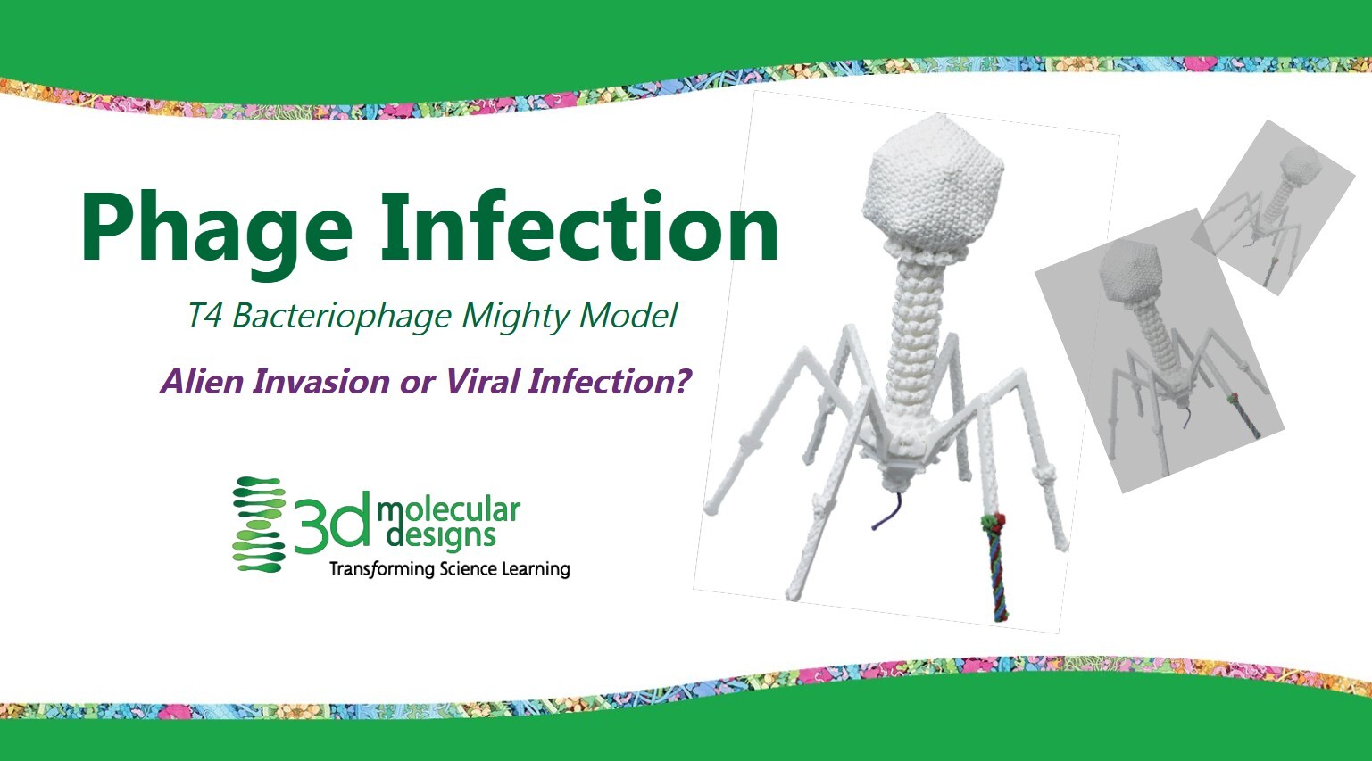
T4 Bacteriophage Mighty Model©Your students will explore the infection process of T4 bacteriophage viruses as they interact with E. coli bacteria in this hands-on, inquiry-driven lesson. Through modeling with the T4 Bacteriophage Mighty Model and a detailed graphic of the E. coli cell wall, students will physically and visually investigate the virus's structure and function and its bacterial host. Through guided observation, students compare and contrast the phage and bacterial membrane, make predictions about the infection process, and use augmented reality features to examine the phage structure further. This activity is highly interactive, incorporating individual reflection, group discussions, and creative modeling. Students are prompted to record observations, sketch and label biological structures, and predict the four-step infection process: bacteria recognition, membrane binding, membrane penetration, and DNA injection. The activity aligns with Next Generation Science Standards, emphasizing model development, explanation construction, and understanding of biological structure and function. Teacher notes and student prompts are embedded throughout, supporting effective facilitation and encouraging student engagement with core concepts in microbiology and virology.
-
Bacteriophage Mighty Model© Replication & Release Slide Deck
![]() Associated Product:
Associated Product:
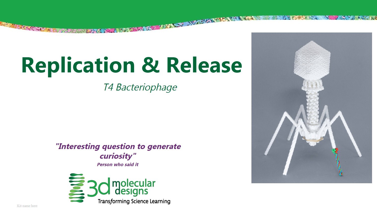
T4 Bacteriophage Mighty Model©Your students will explore the life cycle of T4 bacteriophage viruses as they interact with E. coli bacteria in this hands-on, inquiry-driven lesson. Through modeling with the T4 Bacteriophage Mighty Model and David Goodsell’s T4 Bacteriophage graphic, students will physically and visually investigate the virus's structure and function as it co-opts its bacterial host. This activity is highly informative with embedded videos, and incorporates individual reflection, group discussions, and creative modeling. Students are prompted to record observations, sketch and label biological structures, and make predictions. The activity aligns with Next Generation Science Standards, emphasizing model development, explanation construction, and understanding of biological structure and function. Teacher notes and student prompts are embedded throughout, supporting effective facilitation and encouraging student engagement with core concepts in microbiology and virology. and virology.
-
Aquaporin Exploratorium

![]() Associated Product:
Associated Product:
Mighty Model Channel Protein Collection©Students can easily explore, manipulate, and model the Aquaporin protein in our web-based protein visualization tool. By applying their knowledge of primary, secondary, and tertiary protein structure, students determine the structure and function of the water channel. Aquaporin selectivity and inhibition by mercury are also explored.
-
Flow of Genetic Information Kit© Student Handouts and Teacher Guides
![]() Associated Product:
Associated Product:
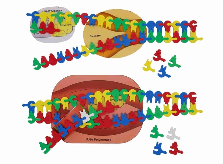
Flow of Genetic Information Kit©The Flow of Genetic Information Kit© allows students to:
- Identify essential enzymes like helicase and polymerase
- Model replication of the leading and lagging strands of DNA
- Explore transcription as they copy one strand of DNA into mRNA using an RNA polymerase
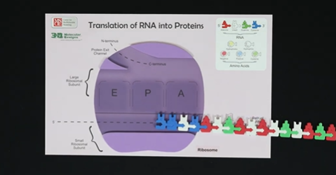
- Engage in translation/protein synthesis as they decode the mRNA into protein on the ribosome placemat
- Reenact the different results of the Meselson and Stahl experiments
-
Phospholipid & Membrane Transport Kit© Student Handouts and Teacher Guides
![]() Associated Product:
Associated Product:
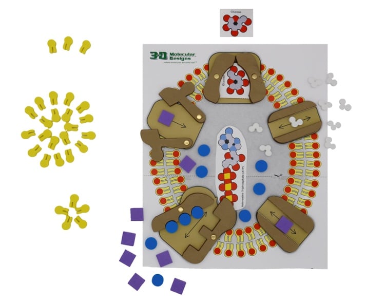
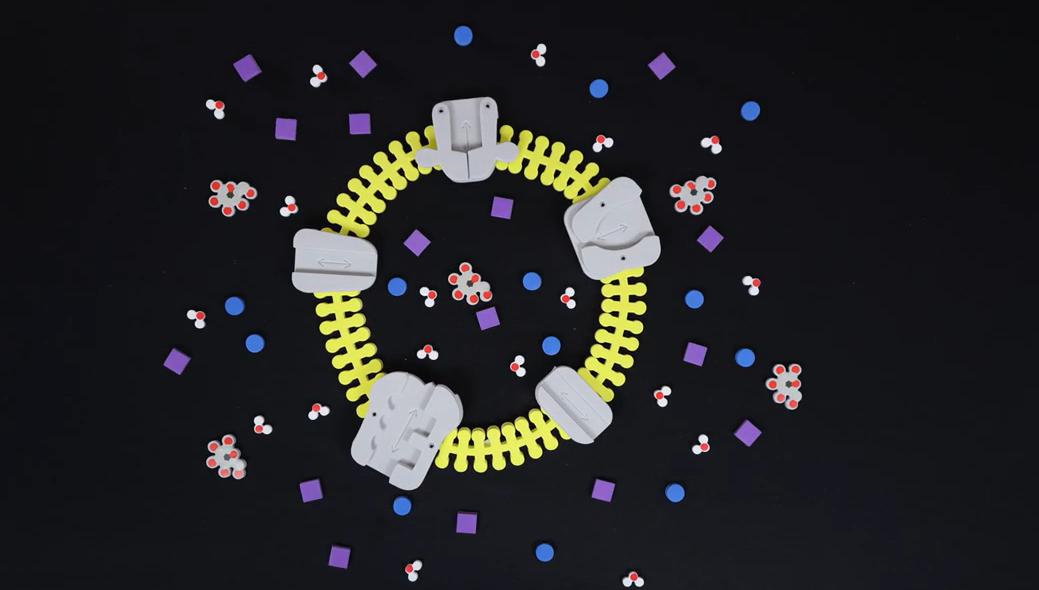
Phospholipid & Membrane Transport Kit©Students learn how about the structure and function of phospholipids, how cell membranes form, and how water and ions move in and out of cells via transport proteins. Including:
- 50 individual phospholipid molecule models demonstrate hydrophobic and hydrophilic concepts to create monolayers, micelles, and bilayers
- Water molecule models show polarity and how water interacts with cell membranes
- Bilayer membrane model creates a cell structure that is flexible yet sturdy
- Active and passive transport with 5 different proteins, ion models, and ATP
Students can model different conditions and then predict which way water will move, making vocabulary like hypertonic, hypotonic, and isotonic more memorable.
-
DNA Monomer to Polymer Interactable
-
Comparing Phospholipid Monolayers, Micelles, and Bilayers Short Content Video

![]() Associated Product:
Associated Product:
Phospholipid & Membrane Transport Kit©Ruth Hutson models phospholipid structure and explores phospholipids' unique behavior in water. Learners are encouraged to model along by creating a monolayer, micelle, and lipid bilayer. Learners are challenged to apply their knowledge of chemical properties of phospholipids and proteins to create a membrane that includes integral proteins.
4:02 Video Length
-
Water Monomer to Polymer Interactable
![]() Associated Product:
Associated Product:
Water Kit©Students build their understanding of the structure of water through digital modeling in this web-based Interactable. Using three different representations of water, students model how atomic interactions within the water molecule help it form hydrogen bonds with twelve other water molecules to make an ice lattice.
-
Side Chain Explorer AR

![]() Associated Product:
Associated Product:
Amino Acid Starter Kit©Students can explore the structure of amino acids and how interactions with water and other amino acids drive protein folding. By applying one of four overlays, students can discover how amino acids interact with each other, evaluate amino acid structure, and classify them by their chemical properties. Based on the data they collect, they can determine the rules of protein folding.
-
A Neighborhood of Cells Slide Deck
![]() Associated Product:
Associated Product:
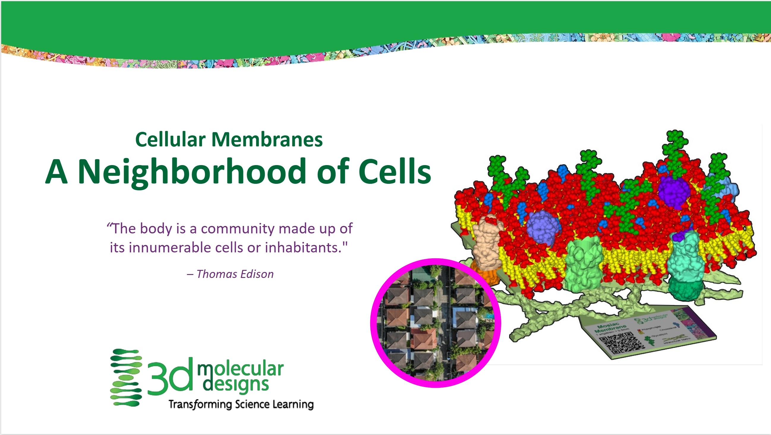
Mosaic Membrane Mighty Model©Students learn about cell membranes by modeling with the Mosaic Membrane Mighty Model and comparing biological membranes to a house. Through modeling and this metaphor, students learn about the flexible and fluid nature of membranes. In addition, they discover how the embedded molecules in a membrane help regulate the rigidity of the membrane, the passage of polar molecules across a membrane, identification, and cell communication.
-
Designing a Biological Membrane Slide Deck
![]() Associated Product:
Associated Product:
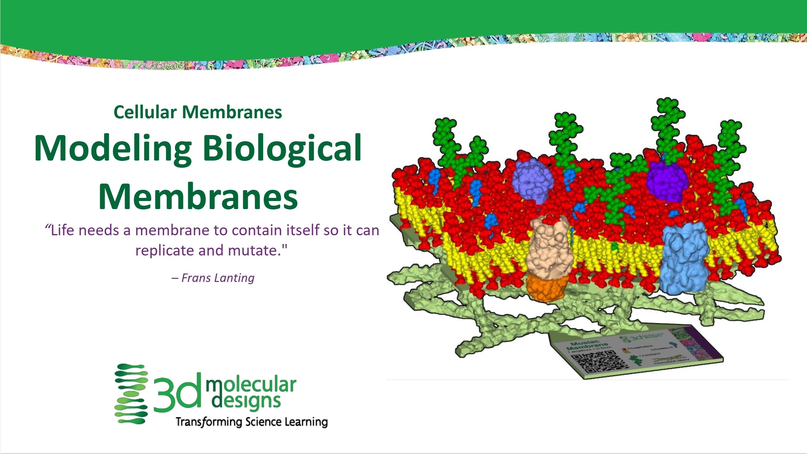
Mosaic Membrane Mighty Model©In this activity, you can invite your students to engage with a tactile membrane model that will help them understand the basic properties, structures and functions of a biological membrane in cells. They will design their own membrane and explore how changing the membrane components will alter the functions supported by their membrane. They will evaluate the limitations of the model and apply their knowledge to make claims about real cell types and membrane protein functions.
-
Side Chain Properties Interactable
![]() Associated Product:
Associated Product:
Amino Acid Starter Kit©Students determine the rules of tertiary protein folding. Amino acid identification and sorting by property, amino acid interactions, and generalized locations of amino acids in the final three-dimensional shape of the protein are explored through this interactive digital medium.
-
Comparing Purines and Pyrimidines Short Content Video
-
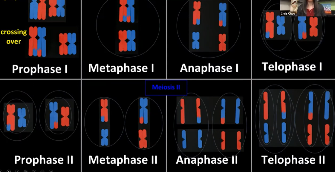
Meiosis Video
-
Water Kit© Overview
![]() Associated Product:
Associated Product:
Water Kit©Learn about a tool to help you teach water concepts, with 3DMD Partner and Director of Operations Heather Ryan, Students use these magnetic water molecules to discover hydrogen bonding, make ice, dissolve salt, evaporate water, explore transpiration, create ethanol, and much more
-
Evolution in Action: Modeling Insecticide Resistance in Mosquitoes
![]() Associated Product:
Associated Product:
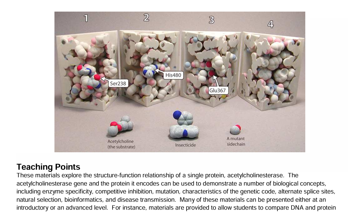
Acetylcholinesterase Active Site Mighty Model©These materials explore the structure-function relationship of a single protein, acetylcholinesterase. The acetylcholinesterase gene and the protein it encodes can be used to demonstrate a number of biological concepts, including enzyme specificity, competitive inhibition, mutation, characteristics of the genetic code, alternate splice sites, natural selection, bioinformatics, and disease transmission. Developed by the MSOE Center for BioMolecular Modeling.
-
Biotechnology Kit© Resources Student Handouts and Teacher Guides
![]() Associated Product:
Associated Product:
Biotechnology Kit©Model diagnostics, forensics, and bacterial transformation at the molecular level. Using the color-coded foam nucleotides, students can:
- Simulate PCR using flanking primers result to copy sequences of interest
- Investigate how restriction endonucleases are used in DNA cloning
- Explore the Sanger DNA sequencing method using chain-terminating fluorescent dideoxy nucleotide analogs
-
States of Matter Short Content Video
-
Amino Acid Starter Kit© Student Handouts and Teacher Guides
-
Acetylcholinesterase Active Site Activity Slide Decks
![]() Associated Product:
Associated Product:
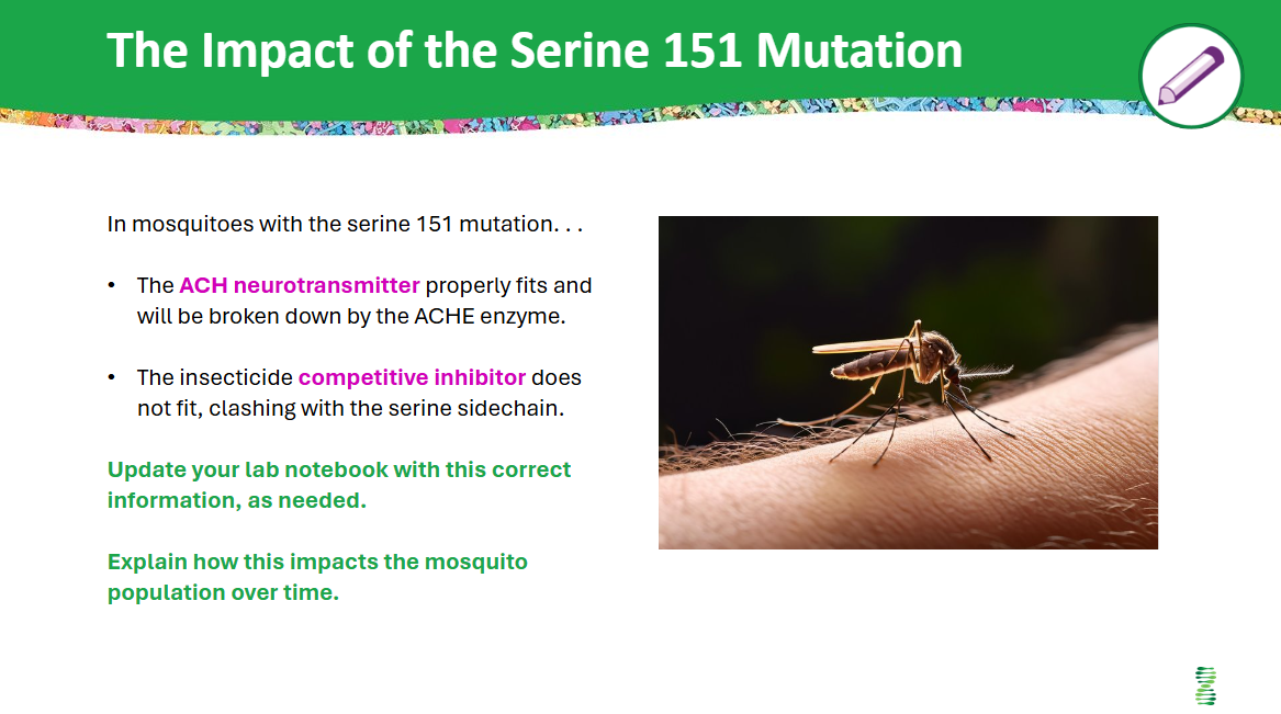
Acetylcholinesterase Active Site Mighty Model©Your students will learn about neurotransmitters, enzyme active sites, competitive inhibitors, mutations, genetic diversity, and evolution through the lens of mosquito resistance to insecticides with the Acetylcholinesterase (ACHE) Active Site Model.
The slide deck will support your students as they generate questions, make predictions and test those predictions with their interactive physical models.
-
Building a Phospholipid Short Content Video
![]() Associated Product:
Associated Product:
Phospholipid & Membrane Transport Kit©Ruth Hutson models a dehydration synthesis reaction to create phospholipids. Learners consider the differences between trans and cis configurations and compare several types of models used to construct membranes. Learners are challenged to model a membrane of their own design.
3:43 Video Length
-
Zinc Finger Exploratorium
![]() Associated Product:
Associated Product:
Amino Acid Starter Kit©Students can easily explore, manipulate, and model the Zinc Finger motif in our web-based protein visualization tool. By applying their knowledge of primary, secondary, and tertiary protein structure, students determine the structure and function of Zinc Fingers.
-
Anabolic Reactions Interactable
![]() Associated Product:
Associated Product:
Enzymes in Action Kit©Students build their understanding of anabolic reactions through digital modeling in this web-based Interactable. Activation energy with and without enzymes is explored. Students also examine active sites and compare glycogen synthase with the schematic model.
-
Chromosome Connections Kit© Student Handouts and Teacher Guides
![]() Associated Product:
Associated Product:
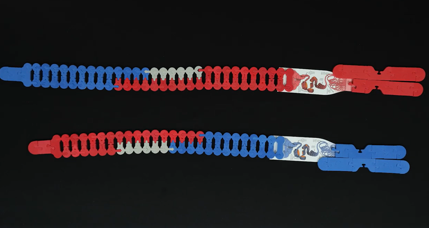
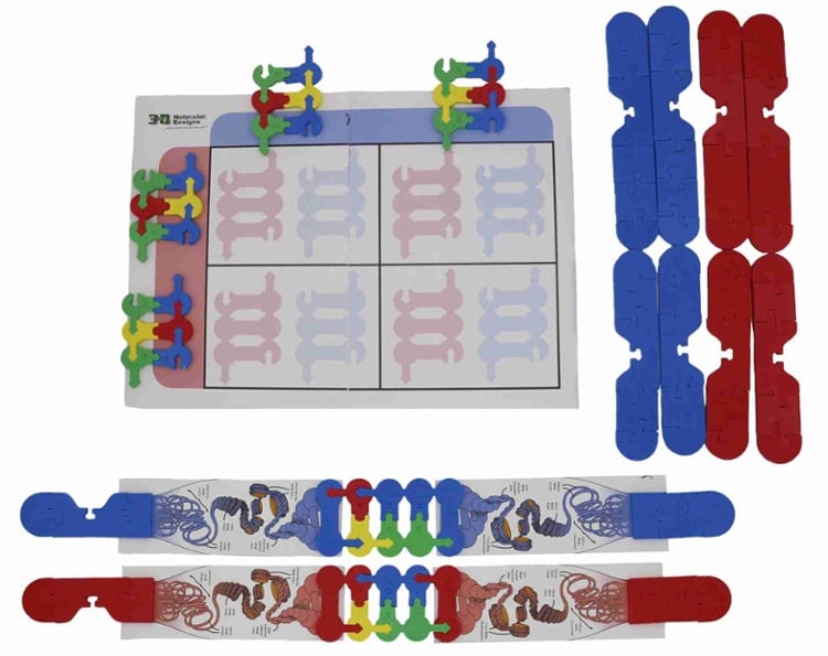
Chromosome Connections Kit©Students to investigate chromosomes at the microscopic and molecular scales. Students learn about the cell cycle, cell division, and genetics. Students will:
- Build homologous chromosome pairs and sister chromatids
- Identify chromosome structure and anatomy
- Construct Punnett squares with gene sequences as alleles
- Compare and contrast mitosis and meiosis
- Explore how genes can cross over during prophase
- Investigate chromosomal nondisjunctions, translocations, inversions, deletions, and duplications
-
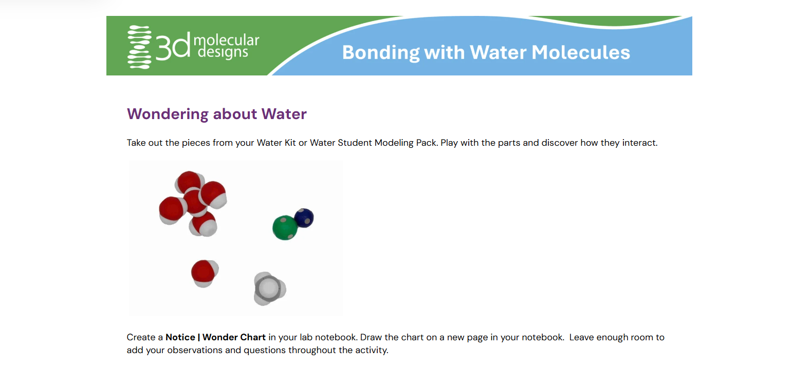
Bonding with Water Molecules Introductory Water Student Modeling Pack Lesson
-
Binding Chemical Interactable
-
Transferring Between Models with the Flow of Genetic Information Kit AR
-
Binding Shape Interactable
-
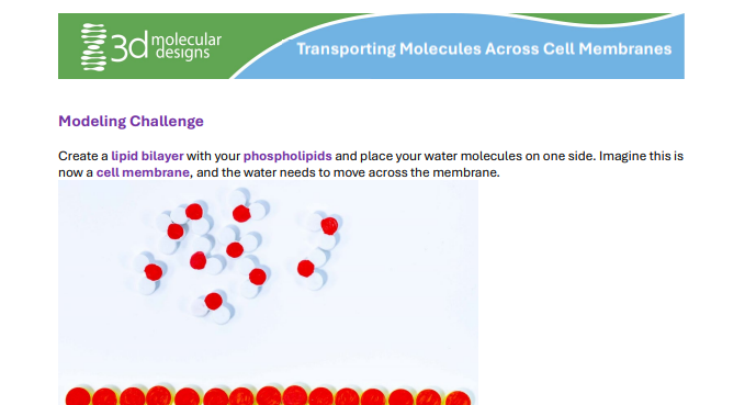
Active & Passive Transport Introductory Membrane Student Modeling Pack Lesson
-
ß-Globin Folding Kit© Student Handouts and Teacher Guides
![]() Associated Product:
Associated Product:
ß-Globin Folding Kit©Reveal the molecular relationship between structure and function, protein synthesis, and mutation as you tell classic stories in biology. Oxygen transport and how it can be disrupted becomes real in your students' hands as they interpret folding maps to form ß-globin. You can create an authentic application activity as students predict the impact of a single-point mutation on the function of the protein. This kit is most effective after students have explored the Amino Acid Starter Kit©.
In biology, oxygen transport and how it can be disrupted becomes real in your students' hands as they:
- Interpret folding maps to form the primary structure of the protein with mini toobers
- Fold 3 mini toobers into alpha helices and then their tertiary structures
- Assemble the three fragments together to form ß-globin
- Add the heme group with an iron atom and O2
-
Passive Transport Stop Motion
-
Amino Acid Starter Kit© Overview
![]() Associated Product:
Associated Product:
Amino Acid Starter Kit©Tim Herman, PhD, walks through the Amino Acid Starter Kit, an engaging, hands-on protein folding kit that allows students to fold a protein while exploring how the chemical properties of amino acids determine its final structure. Dr. Herman is founding partner and scientific officer at 3D Molecular Designs. He is founder and director of the MSOE Center for BioMolecular Modeling.
-
Transferring Between Models with the Dynamic DNA Kit AR
-
Protein Structure Modeling Kit Activity Slide Deck
![]() Associated Product:
Associated Product:
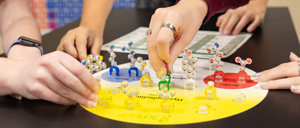
Protein Structure Modeling KitStudents learn the structure and function of proteins by building a physical model of a specific protein of their choice. As they design their model, students will learn the levels of protein structure—primary, secondary, and tertiary.
Students will create their models from 3-dimensional computer renderings of a protein. Options for creating these computer visualizations of their protein are the Protein Exploratorium or JUDE (Jmol User Design Environment). Videos of how to fold a Modeling Tie are included.
-
Exploring the Solubility of Alcohols Video
-
Building a Triglyceride Short Content Video
-
3DMD Jmol Training Guide
![]() Associated Product:
Associated Product:
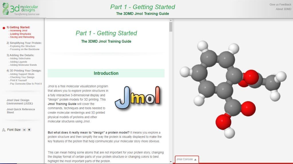
SMART Team ProgramThis free, four part web series includes all of the information and training needed to access the molecular visualization program Jmol, design a unique protein model, and export that model for 3D printing. Jmol is used extensively in 3DMD's SMART Team program, Science Olympiad Protein Modeling event, and 3D printing training resources.
-
A Tale of Two Sisters: Modeling Transcription and Translation in Protein Synthesis
![]() Associated Product:
Associated Product:
Nucleotide Student Modeling Pack©Fran Grant of Cedarburg High School (Cedarburg, WI) and Mark Arnholt of Hartford Union High School (Hartford, WI) explore using 3DMD’s Flow of Genetic Information Kit and Nucleotide Student Modeling Pack to model protein synthesis with students, whether they are learning virtually or in the classroom. They also consider the story of the Obando sisters and their cutting-edge treatments for sickle cell disease.
-
Phospholipids Introductory Membrane Student Modeling Pack Lesson
-
Making the Cut with CRISPR-Cas9 Kit© Resources Student Handouts and Teacher Guides
![]() Associated Product:
Associated Product:
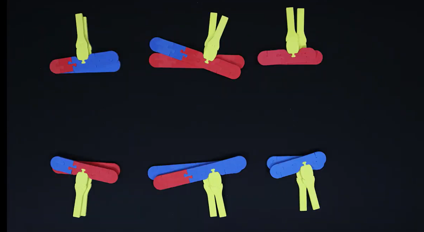
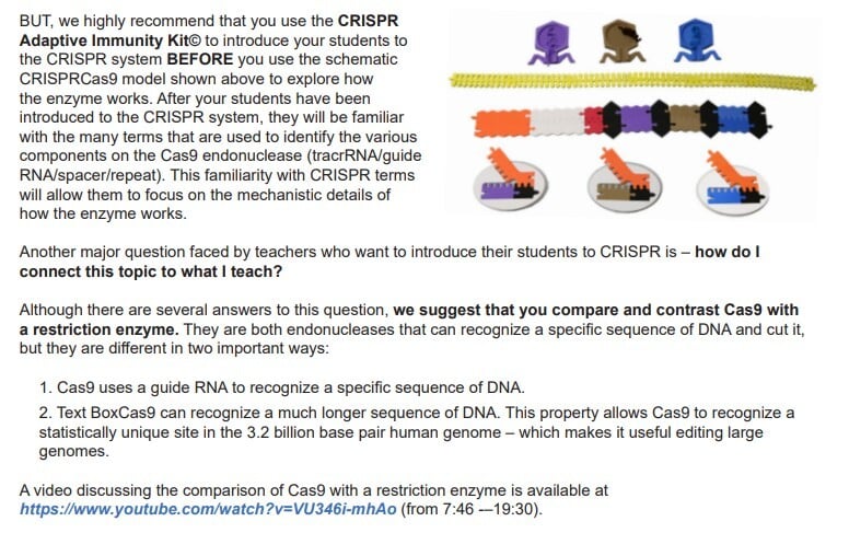
Making the Cut with CRISPR-Cas9 Kit©An effective search-and-destroy CRISPR system in bacteria-inspired innovation in genetic technology. Your classroom can become a design solutions lab as students discover the role of the Cas9 protein in defending against bacteriophages. With engaging foam pieces and chenille bacteriophage genomes, your students can model the natural process that inspired genetic engineering innovations.
-
CRISPR Adaptive Immunity Kit© Resources Student Handouts and Teacher Guides
![]() Associated Product:
Associated Product:
CRISPR Adaptive Immunity Kit©An effective search-and-destroy CRISPR system in bacteria-inspired innovation in genetic technology. Your classroom can become a design solutions lab as students discover the role of the Cas9 protein in defending against bacteriophages. With engaging foam pieces and chenille bacteriophage genomes, your students can model the natural process that inspired genetic engineering innovations.
-
Protein Structure Tutorials
![]() Associated Product:
Associated Product:
Protein Structure Modeling KitStudents will learn the four levels of protein structure—primary, Secondary, Tertiary, and quaternary—through interactive web pages and embedded videos. The tutorial includes examples of specific proteins, and students will compare and manipulate multiple representations as they build their knowledge through the levels of protein structure.
-
Exploring States of Matter AR
![]() Associated Product:
Associated Product:
Water Kit©Students can model water molecule behavior by exploring how molecular water exists as a solid, liquid, or gas, depending on temperature. Using two scenes, either a single water molecule or a six-molecule ice lattice is scanned. By increasing temperature using a slider bar, students can increase the average energy of motion of the water molecules, allowing hydrogen bonds between them to break. Students can also explore and calculate density of the states of matter.
-
Components of a Phospholipid Short Content Video
![]() Associated Product:
Associated Product:
Phospholipid & Membrane Transport Kit©Ruth Hutson explores the diversity of phospholipids in a cell membrane. Learners are encouraged to model four phospholipids and determine their similarities and differences. Learners are challenged to make a lipid bilayer and explore the dynamic nature of this structure.
4:13 Video Length
-
Neuromuscular Synapse Poster© Student Handouts and Teacher Guides
![]() Associated Product:
Associated Product:
Neuromuscular Synapse Poster©Glimpse how the hundred trillion cells in our brains make things happen. This captivating watercolor poster leads students through the molecular interactions found at neuromuscular synapses at one million times magnification.
This unique watercolor image created by David S. Goodsell, Ph.D., connects the molecular world, inferred by X-ray crystallography and NMR spectroscopy, with the cellular world, observed by light and electron microscopy.
- Explore the connection between a nerve cell and a muscle
- Examine vesicles filled with the neurotransmitter acetylcholine fuse with the membrane of the pre-synaptic axon
- Follow acetylcholine, which diffuses across the synaptic space to bind to acetylcholine receptors of the muscle cell
- Discover acetylcholinesterase entangled in the synaptic space, where it breaks down excess acetylcholine
David S. Goodsell, Ph.D., scientist, author, and artist of all things small, creates cellular landscapes that accurately illustrate the size, shape, and distribution of proteins in their natural cell environment. These unique watercolor images connect the molecular world, inferred by X-ray crystallography and NMR spectroscopy, with the cellular world, observed by light and electron microscopy.
Learn more about the Neuromuscular Synapse Poster©
The Neuromuscular Synapse Poster measures 23" x 30".
-
Side Chain & Protein Property Interactable
-
Catabolic Reactions Interactable
-
AR Training App Resources
![]() Associated Product:
Associated Product:
Amino Acid Starter Kit©
Dynamic DNA Kit©
Flow Of Genetic Information Kit©
Water Kit©The 3DMD AR app allows students to further explore 3D Molecular Designs' kits and models through augmented reality-based activities. 3DMD AR uses physical models as AR targets and leads students through digital learning activities. As students progress through AR activities, they will continue to manipulate physical models, actively engaging in making sense of molecular processes through collaboration with classmates. Additional resources are available to help students learn to navigate the app.
-
CRISPR-Cas9 System Video
![]() Associated Product:
Associated Product:
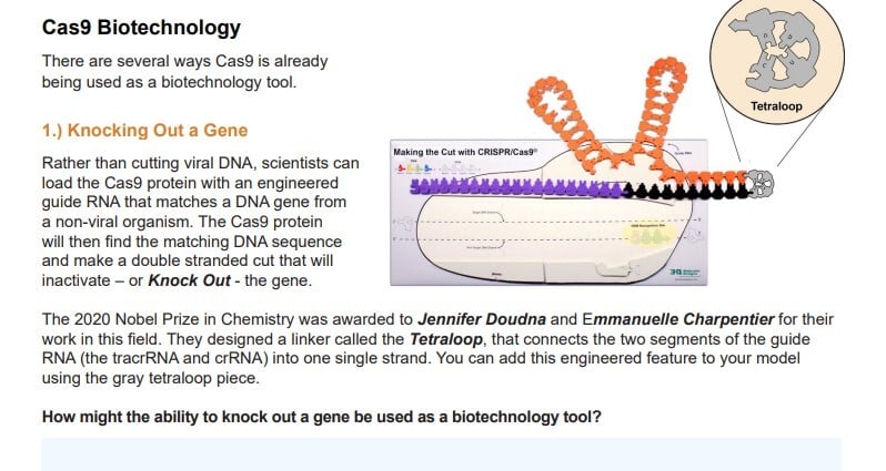
Making the Cut with CRISPR-Cas9©Your students have heard about CRISPR, now, they can model how this protein can modify DNA. With Making the Cut with CRISPR-Cas9©'s colorful foam pieces and placemat your students can build an interactive model of the CRISPR-Cas9 RNA-guided endonuclease, including the viral double-stranded DNA and dual guide RNA. You can observe students transferring their prior knowledge of DNA structure while modeling how the Cas9 protein functions.
- Build a schematic model of the CRISPR-Cas9 RNA-guided endonuclease, including the viral double-stranded DNA and dual-guide RNA
- Understand how the Cas9 protein binds to a specific sequence of viral double-stranded DNA at a PAM site - and cuts it
- Demonstrate how that cut destroys the viral genome and prevents the infection of bacteria
- Amplify their learning with student handouts and digital materials
You can observe students transferring their prior knowledge of DNA structure while modeling how the Cas9 protein functions.
-
Protein Translation with the Flow of Genetic Information Kit AR
![]() Associated Product:
Associated Product:
Flow of Genetic Information Kit©Students explore the process of protein synthesis and how certain antibiotics block that process. Using 5 scenes, students review the steps in protein translation and then predict how four antibiotics interact with different ribosome locations and disrupt protein formation.
-
Flow of Genetic Information Kit© Demonstration Video
![]() Associated Product:
Associated Product:
Flow of Genetic Information Kit©3DMD Partner and Director of Operations, Heather Ryan, explores how students can model replication of the leading and lagging strands of DNA, model transcription as they copy one strand of DNA into mRNA using an RNA polymerase, and model translation/protein synthesis as they decode the mRNA into protein on the ribosome placemat.
-
Discovering Polarity AR
![]() Associated Product:
Associated Product:
Water Kit©Students explore electron distribution in a water molecule to determine the role of the electron in covalent bonding. Using three scenes, students conclude that electrons are not equally shared, resulting in partial positive and partial negative regions on the water molecule. They explore the unequal charge across the water molecule to further understand water molecule polarity and hydrogen bonding.
-
Folding Real Proteins-Zinc Fingers AR
![]() Associated Product:
Associated Product:
Amino Acid Starter Kit©Students can explore the levels of protein structure using four scenes and fold a zinc finger as a challenge. They will determine that the sequence of amino acids makes a protein's distinctive primary sequence. Secondary structure gives a protein greater stability and is made of alpha helices and beta sheets. By folding a real protein, students will notice the tertiary structure is the final overall structure. Students will interact a zinc finger with DNA to discover its structure allows it to perform a unique function.
-
Translation Stop Motion
-
Tour of a Human Cell© AR
![]() Associated Product:
Associated Product:
Tour of a Human Cell©Invite students to journey from the nucleus to the outer cell membrane with this beautiful watercolor poster by scientist and artist David Goodsell, a Ph.D. example of the immunology of your flu shot. printed by 3D Molecular Designs. This unique image connects the molecular world, inferred by X-ray crystallography and NMR spectroscopy, with the cellular world, observed by light and electron microscopy.
Printed by 3D Molecular Designs.
The Panorama© and Grand Panorama© capture students' imaginations as they:
- Identify how DNA is wrapped around histones forming nucleosomes
- Explore RNA polymerase unwrapping the DNA and making mRNA
- Examine mRNA moving through nuclear pore complexes to ribosomes, the site of protein production
- Discover antibody proteins delivered into the endoplasmic reticulum
- Observe vesicles carrying these proteins through the Golgi
- Notice the kinesin motor proteins pulling the antibodies to the cell membrane to be released
Captivating shapes and colors tell the story of cell organelles working as a system to prepare antibodies as an immune response.
Dr. Goodsell creates cellular landscapes that accurately illustrate the size, shape, and distribution of proteins in the cells. These unique images connect the molecular world, inferred by X-ray crystallography and NMR spectroscopy, with the cellular world, observed by light and electron microscopy.
-
Molecule Maker Online
-
Physical and Chemical Properties of Water
-
Extending the DNA Double Helix AR
![]() Associated Product:
Associated Product:
Dynamic DNA Kit©Students explore the structure of DNA and how other molecules interact with DNA. Throughout four scenes, they extend the DNA double helix to view the major and minor grooves, look at how nucleosomes help store and organize DNA, determine how RNA polymerase makes transcripts of genes, and how transcription factors help regulate access to DNA.
-
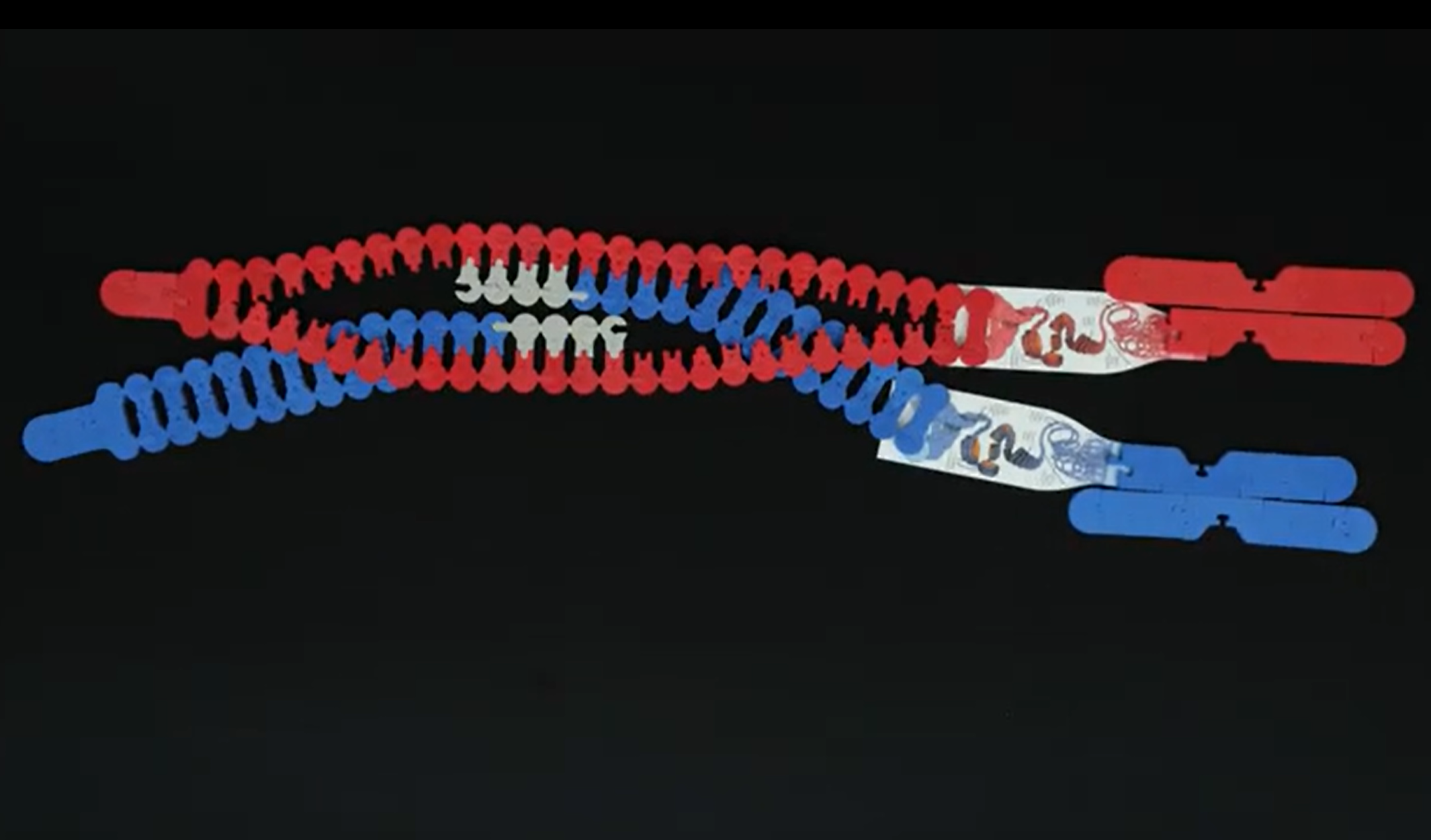
Making the Cut with CRISPR Cas-9 Webinar
-
Enzymes in Action Kit© Student Handouts and Teacher Guides
![]() Associated Product:
Associated Product:
Enzymes in Action Kit©Models give words meaning. These colorful foam pieces with intuitive shapes let your students experience enzymes in action before they even know any vocabulary. Collaboration and discourse naturally occur between learners as they figure out what it means for a protein to be a catalyst. Excellent at revealing student thinking, this kit can complement traditional classroom assignments like the catalase or amylase labs.
-
Water Kit© Student Handouts and Teacher Guides
![]() Associated Product:
Associated Product:
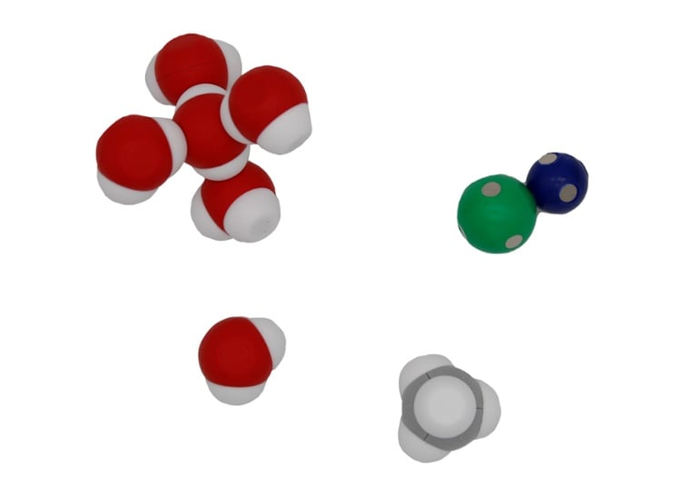
Water Kit©Students learn about molecular attractions and repulsions, providing a springboard to more complex chemistry concepts. When using this kit, students can:
- Investigate the polarity of water molecules
- Explore ionic, covalent, and hydrogen bonds
- Experiment with adhesion, cohesion, and capillary action
- Identify and model states of matter
- Demonstrate changes in volume between liquid and solid water
- Build different types of ice
- Simulate solubility with sodium and chloride ions
- Build ethane and ethanol to observe interactions with water
-
Crossing over - molecular scale
-
Keep Your Head Above Water: Constructing and Crossing Cell Membranes
![]() Associated Product:
Associated Product:
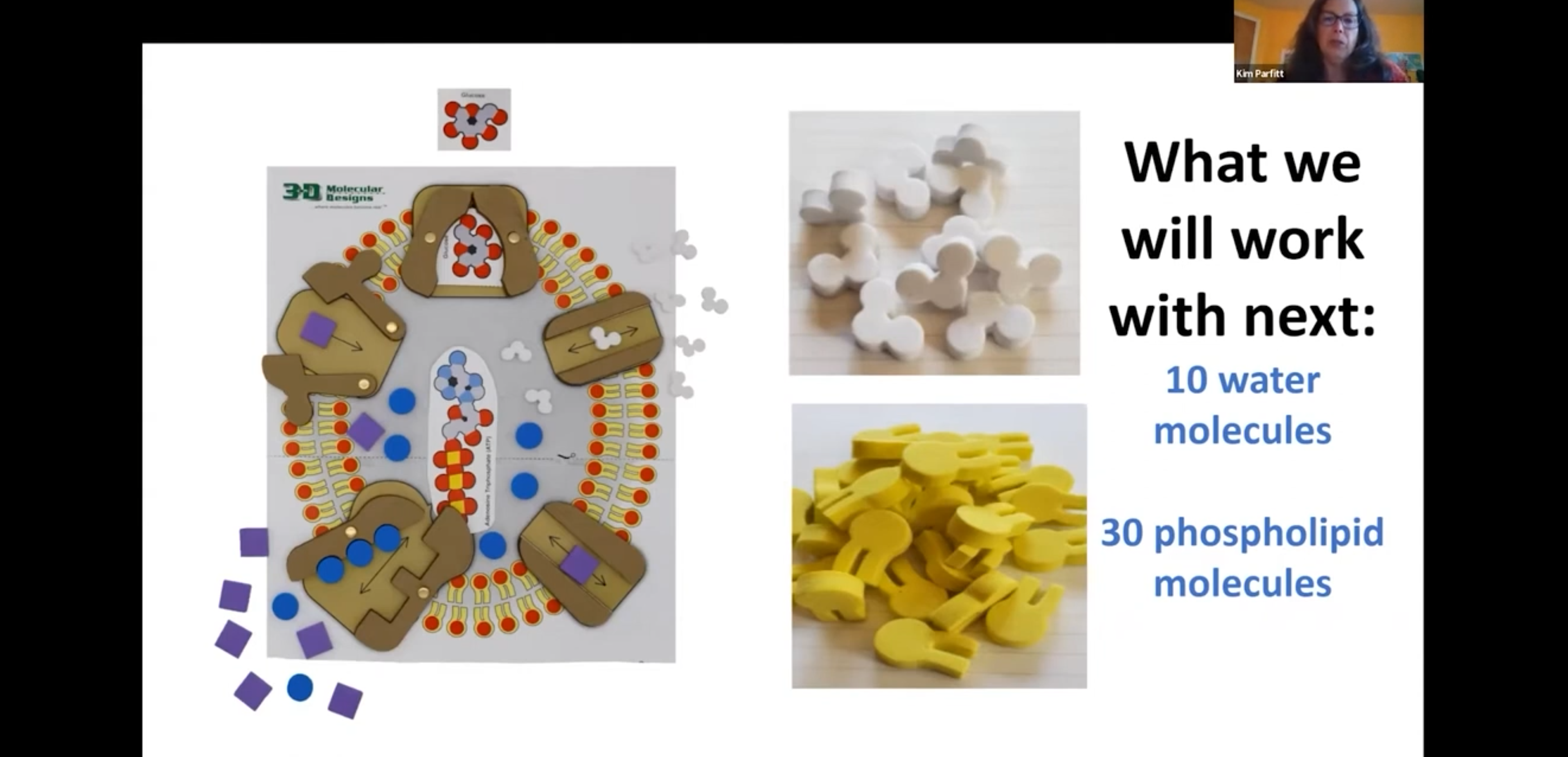
Membrane Student Modeling Pack©Science educator Kim Parfitt uses 3D Molecular Designs' Phospholipid & Membrane Transport Kit to demonstrate how students can explore the beautiful world of membranes while predicting, revising and justifying their own models. Looking for patterns in nature will reveal how these structures enable the functions for life to survive at different scales.
-
Insulin mRNA to Protein Kit© Student Handouts and Teacher Guides
-
Chromosomes and DNA Webinar
-
Unfolding the Mystery of Protein Structure
![]() Associated Product:
Associated Product:
Protein Student Modeling Pack©Ready to fold your way through the basic chemistry of amino acids and the complex structure of proteins? Jill Daniels of the Geneva School of Boerne (Boerne, TX) explores an easy-to-follow hands-on activity from 3D Molecular Designs that will become a must have for your teacher arsenal. You will be amazed how adding modeling to this mystery will unfold protein complexity!
-
NaCl Lattice© Student Handouts and Teacher Guides
![]() Associated Product:
Associated Product:
NaCl Lattice©With this model, students will:
- Discover the cubic nature of salt crystals
- Explore how the positive sodium and negative chloride ions form electrostatic bonds
- Examine the efficient lattice packing of the ions
- Investigate high melting temperature, brittleness, and cleavage planes
-
DNA Starter Kit© Resources Student Handouts and Teacher Guides
![]() Associated Product:
Associated Product:
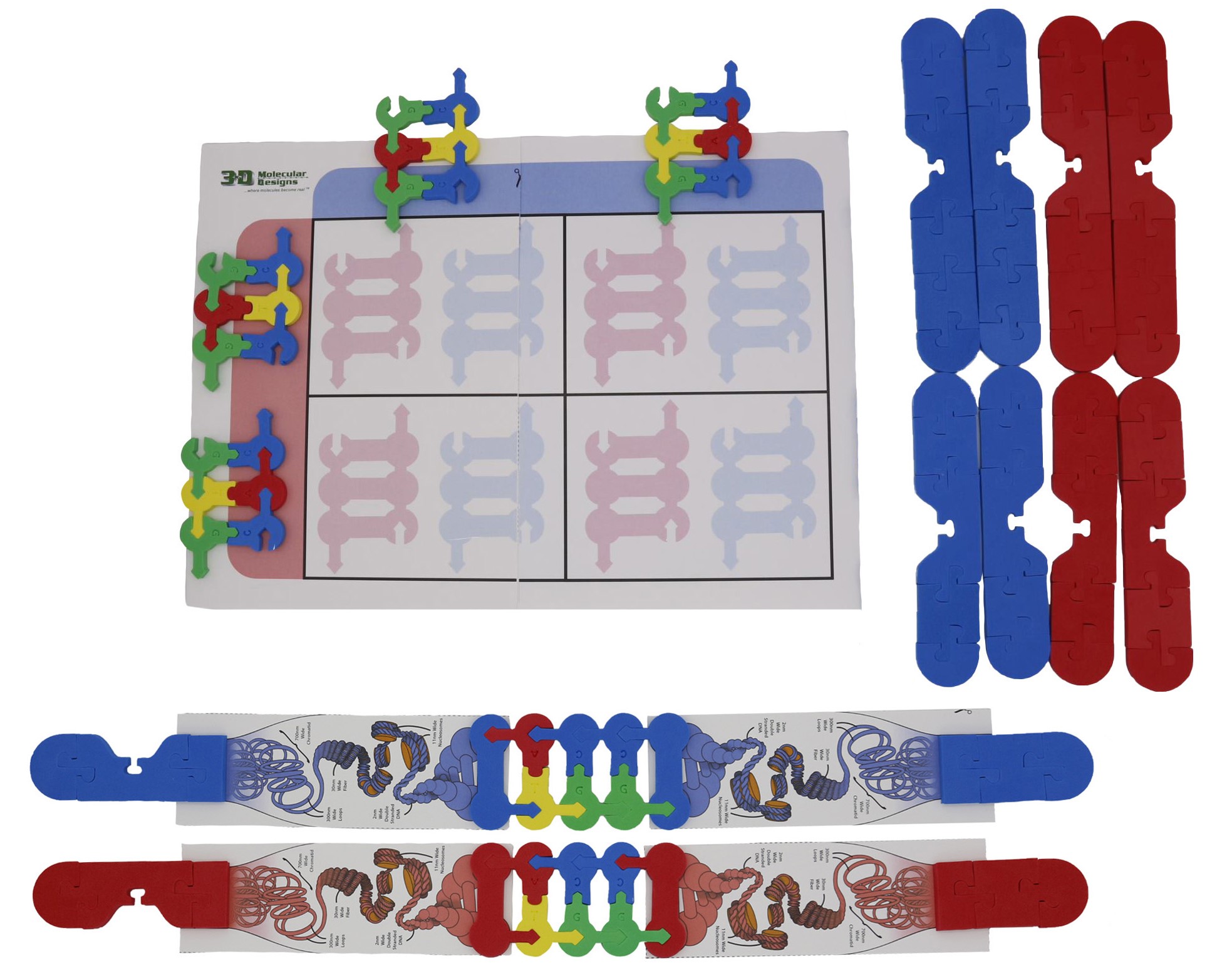
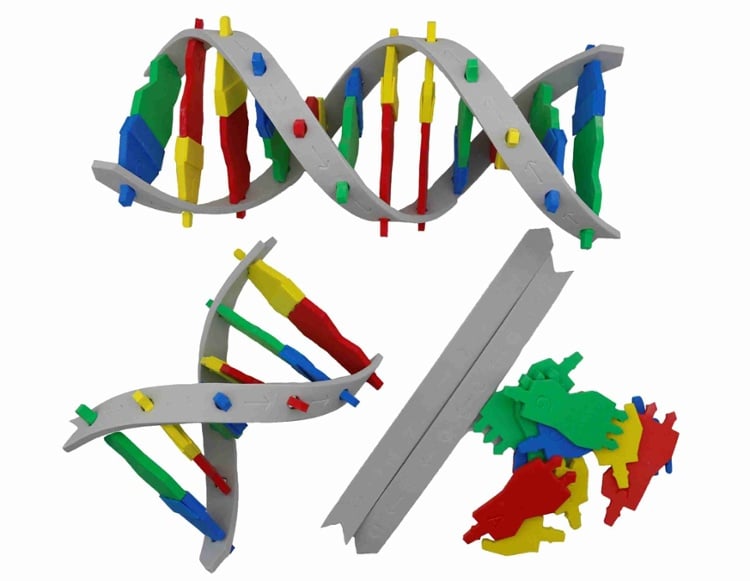
DNA Starter Kit©3D Molecular Designs' interactive foam DNA model makes double-stranded DNA and single-stranded RNA. Introduce the basics of DNA structure, then move into replication and transcription.
Groups of students can:
- Pair nitrogenous bases using the base pairing rule to create a sequence
- Transform the familiar ladder shape of DNA with a simple twist
- Explore the semi-conservative replication of DNA
- Synthesize mRNA using sugar-phosphate pieces to show transcription
This easy-to-build model is a perfect starting point that students can use to test their ideas and transfer their understanding to other, more complex models.
-
RNA Processing Short Content Video
-
Insulin Animation
-
Chromosome and Gene Structures Short Content Video
![]() Associated Product:
Associated Product:
Chromosome Connections Kit©Ruth Hutson considers the relationship between a gene and a chromosome while comparing their relative size, scale, and proportion. Learners delve into chromosome structure and how histones organize and regulate DNA. Learners are challenged to construct a circular prokaryotic chromosome and then compare it with the arrangement of linear eukaryotic chromosomes.
Video Length
4:48
-
JUDE (Jmol User Design Environment)
![]() Associated Product:
Associated Product:
Amino Acid Starter Kit©JUDE (Jmol User Design Environment) is an online version of Jmol that allows students to design their own proteins from the Protein Database in any browser. The files created can be exported in multiple formats, including JPG for presentations or documents and STL to 3D Print.
-
Modeling PCR
-
Protein Exploratorium
-
Dynamic DNA Kit Student Handouts and Teacher Guides
![]() Associated Product:
Associated Product:
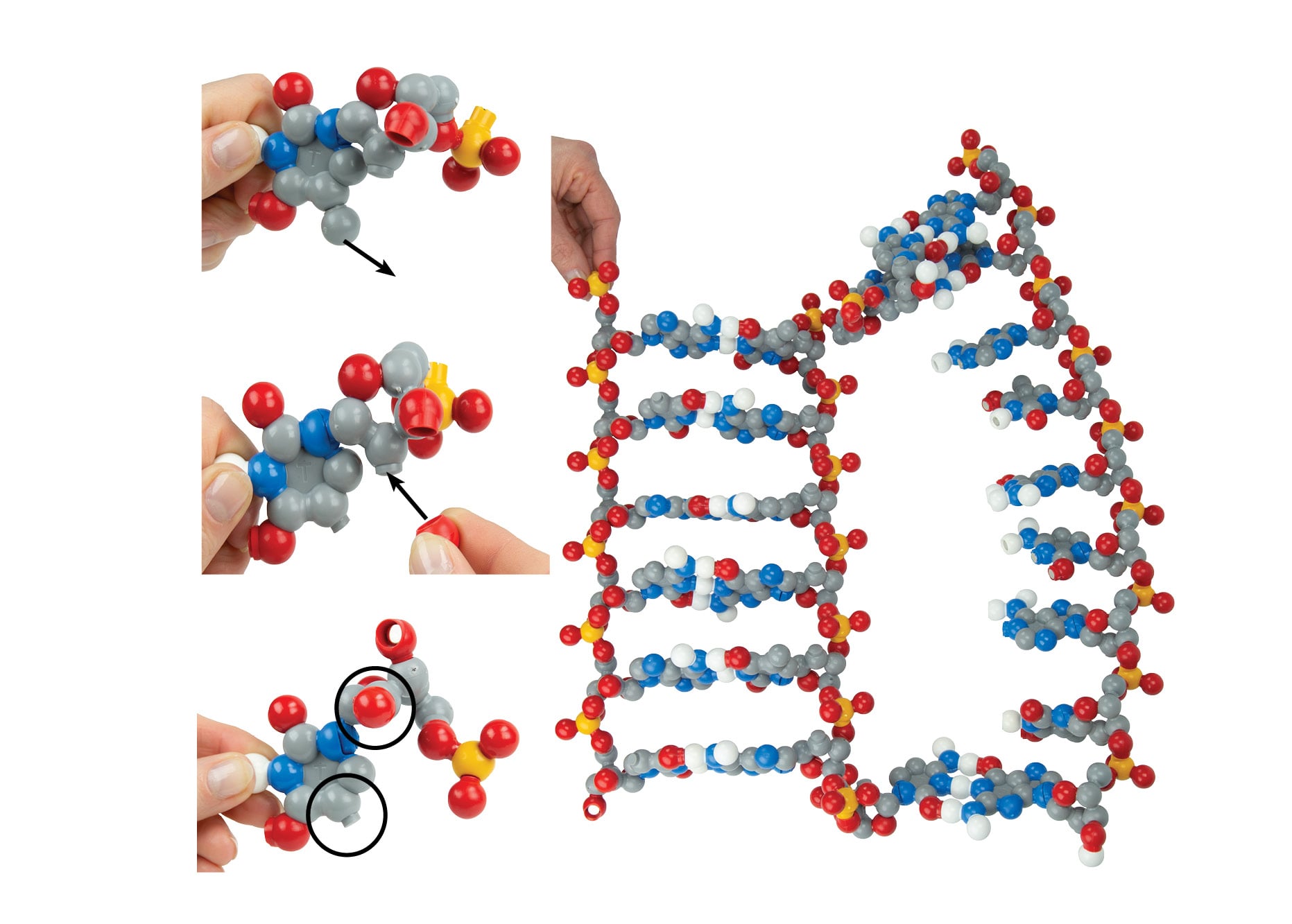
Dynamic DNA Kit©Sturdy, colorful pieces and magnets make it easy for students to snap, bend, twist, and interact as they build DNA. Thanks to our patented design innovations, the structure of this molecule is revealed to students through their own investigation.
- Construct individual nucleotides with nitrogenous bases, sugars, and phosphate groups
- Feel the hydrogen bonding of the A-T and G-C base pairs
- Twist and unwind the double helix to set the stage for replication and transcription
- Convert thymine to uracil
Additional pieces allow them to make ATP, consider epigenetics and gene expression, and much more.
-
Tour of a Human Cell© Student Handouts and Teacher Guides
![]() Associated Product:
Associated Product:
Tour of a Human Cell©Invite students to journey from the nucleus to the outer cell membrane with this beautiful watercolor poster by scientist and artist David Goodsell, a Ph.D. example of the immunology of your flu shot. printed by 3D Molecular Designs. This unique image connects the molecular world, inferred by X-ray crystallography and NMR spectroscopy, with the cellular world, observed by light and electron microscopy.
Printed by 3D Molecular Designs.
The Panorama© and Grand Panorama© capture students' imaginations as they:
- Identify how DNA is wrapped around histones forming nucleosomes
- Explore RNA polymerase unwrapping the DNA and making mRNA
- Examine mRNA moving through nuclear pore complexes to ribosomes, the site of protein production
- Discover antibody proteins delivered into the endoplasmic reticulum
- Observe vesicles carrying these proteins through the Golgi
- Notice the kinesin motor proteins pulling the antibodies to the cell membrane to be released
Captivating shapes and colors tell the story of cell organelles working as a system to prepare antibodies as an immune response.
Dr. Goodsell creates cellular landscapes that accurately illustrate the size, shape, and distribution of proteins in the cells. These unique images connect the molecular world, inferred by X-ray crystallography and NMR spectroscopy, with the cellular world, observed by light and electron microscopy.
-
Folding and Annotating Modeling Ties Short Content Videos
![]() Associated Product:
Associated Product:
Protein Structure Modeling KitStudents will learn how to create a model of a protein using Modeling Ties in the pair of videos. The first video focuses on preparing your Modeling Tie for folding by annotating the primary and secondary structures onto the Tie. In the second video, students are instructed on how to fold the secondary and tertiary structures.
8:10 Video Length
-
Identifying Regions on a Chromosome Short Content Video
![]() Associated Product:
Associated Product:
Chromosome Connections Kit©Ruth Hutson models several important structures found on linear chromosomes and explores how scientists use size, banding pattern, and centromere position to differentiate between them. Learners are challenged to model several different types of chromosomes on their own.
5:52 Video Length
-
Cholinergic Synapse Stop Motion Video
-
Webinar: Chromosomes & DNA - YouTube
![]() Associated Product:
Associated Product:
Chromosome Connections Kit©Conceptual understanding skyrockets as you invite students to investigate chromosomes at the microscopic and molecular scales! You will love the versatility of this kit as you explore the cell cycle, cell division, and genetics. The nucleotides are the same shape and size as those in the Flow of Genetic Information Kit© to provide even more versatility for teaching genetics.
-
Neuron Modeling Kit© Student Handouts and Teacher Guides
-
DNA Structure and Function Webinar
-
Exploring Unsaturated, Saturated, & Supersaturated Solutions Short Content Video
-
Mitosis Video
-
Map Of The Human ß -Globin Gene© Student Handouts and Teacher Guides
![]() Associated Product:
Associated Product:
Map of the Human ß-Globin Gene©Students often wonder if all the DNA in a gene is a code for a protein. Working in teams or small groups, they answer their own question while getting a glimpse of bioinformatics, which is the study of information DNA encodes.
- Initiate their investigation by examining the amino acid sequence of the ß-globin gene
- Locate reading frames
- Utilize triplet codons
- Discover introns and exons
Once students have grasped these eukaryotic gene features, you can examine landmark amino acids, including Glu6 site of a sickle cell mutation, His63 the distal histidine, and His92 is the proximal histidine that binds the heme group. All maps, including the teacher key, are laminated to allow for years of use.
-
Cytochrome C, Immunoglobulin Fold, and Zinc Finger Jmol Explorations
![]() Associated Product:
Associated Product:
Protein Structure Modeling KitTo learn about protein structure and function, students can explore protein structures in Jmol, a molecular visualization tool. They can change the visualization with simple buttons or code more complex commands in the console. The preset proteins are Cytochrome C, an Immunoglobulin Fold, and a Zinc Finger. Links to the PDB file in the Protein Data Bank and the Molecule of the Month entry are included.
-
Substrate Specificity Kit© Student Handouts and Teacher Guides
![]() Associated Product:
Associated Product:
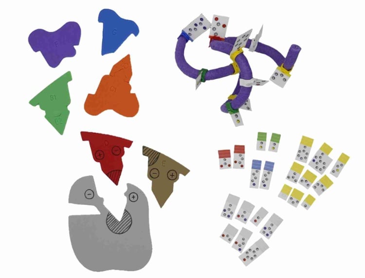
Substrate Specificity Kit©Your students can model the highly specific interaction between a substrate and an enzyme. Students can:
- Build a substrate and identify its chemical properties
- Fold a mini toober to create an active site for that substrate
- Discover subtle changes in enzyme structure that can impact substrate binding
You can even explore the various types of specificity, including stereochemical specificity and absolute specificity.
-
Synapse Construction Kit© Student Handouts and Teacher Guides
![]() Associated Product:
Associated Product:
Synapse Construction Kit©Your students can investigate the action potential of a neuron plus the interaction of neurotransmitters, membrane receptors, and transport proteins. Once students create their model to show the typical process for the neuron function, they can analyze where the process can break down. They then revise their model using representations of nicotine, cocaine, sarin gas, and propofol on neuronal signaling to predict the molecular and organismal response.
-
Modeling Mini Toobers© Student Handouts and Teacher Guides
![]() Associated Product:
Associated Product:
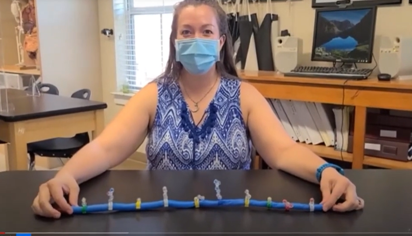
Modeling Mini Toobers©You will want to have these versatile and fun toobers within easy reach throughout the school year. With each kit, students can fold and twist the 1-meter long toober into alpha helices or beta sheets. Use two toobers together to demonstrate the plectonemic nature of DNA. You can easily use these before or after a more thorough exploration of protein folding and structure.
Each flexible foam mini toober has a soft wire core and is 1 meter long. They are sold separately. One red and one blue end cap come with each mini toober. Push pins are not included.
-
Coronavirus Models© Student Handouts and Teacher Guides
![]() Associated Product:
Associated Product:
Coronavirus Models©This evocative SARS-CoV-2 coronavirus model is hard to resist. Your students will immediately want to pick this up - and they can as it is 3D printed using lightweight plastic. Accurate and interactive with embedded magnets, students refer to it while using our FREE Science of Coronaviruses Video Series.
Learn more about the Coronavirus Model©
Model comes with removable base, antibody with a magnet and mini toober RNA viral genome segment.
-
The Data Dilemma© Student Handouts and Teacher Guides
![]() Associated Product:
Associated Product:
The Data Dilemma©"I wonder if" and "What about" will be part of your students' discourse as they think like scientists with this nature of science activity. Students use the geometric shapes to create a single model. As more data becomes available, students revise their thinking, and their models.
- Arrange tangram pieces to represent an initial conceptual model or explanation
- Receive new information that expands or challenges their initial model
- Experiment with multiple solutions to include the new data
- Apply this iterative process to examples of classic scientific advances and theories
A perfect beginning of the school year activity to practice asking questions, working collaboratively, and designing and revising models.
Learn more about The Data Dilemma©
-
Protein Student Modeling Pack© Resources
-
Double Helix Student Modeling Pack© Resources
![]() Associated Product:
Associated Product:
Double Helix Student Modeling Pack©Students can individually explore the basics of DNA structure and replication with these hands-on models. This pack and free digital resources support learning and let students show you their understanding. -Examine the structure of color-coded DNA bases -Identify purines and pyrimidines -Explore hydrogen bonding in A-T and G-C base pairs -Connect bases to a continuous sugar-phosphate backbone -Discover the directionality of each backbone strand -Build double-stranded DNA and twist it into the double helix -Model the process of replication
-
Chromosome Student Modeling Pack© Resources
![]() Associated Product:
Associated Product:
Chromosome Student Modeling Pack©Individual students can spend more time at home or in the classroom to explore chromosomes. Foam pieces help students compare the microscopic and molecular scales of genetic information. This pack with free digital resources supports learning and lets students show you their understanding. -Identify chromosome structure and anatomy -Compare mitosis and meiosis -Discover the sources of genetic variation through independent assortment and crossing over -Construct Punnett squares with nucleotide sequences to reinforce the nature of alleles
-
Membrane Student Modeling Pack© Resources
![]() Associated Product:
Associated Product:
Membrane Student Modeling Pack©Students can individually explore cell membranes and the role of transport proteins in moving ions and small molecules. This pack, along with free digital resources, supports learning and lets students show you their understanding. -Compare and contrast models of phospholipids -Discover the spontaneous formation of cell membranes -Create a micelle and liposome potential for drug delivery -Explore dehydration synthesis reaction in a triglyceride or phospholipid -Identify and simulate the function of proteins involved in membrane transport -Investigate passive and active transport -Simulate how a cell maintains ionic balance Free digital resources support learning and let students show you their understanding.
-
Water Student Modeling Pack© Resources
![]() Associated Product:
Associated Product:
Water Student Modeling Pack©Polarity is unforgettable after modeling with these engaging magnetic molecules. Students love figuring out molecular attractions and repulsions. Providing a springboard to more complex chemistry concepts lets you offer students in home study, in tutoring centers, or in class a chance to work as an individual.
-
Nucleotide Student Modeling Pack© Resources
![]() Associated Product:
Associated Product:
Nucleotide Student Modeling Pack©Students can individually model replication, transcription, and even biotechnology concepts with these models and digital activities. Our Nucleotide Student Modeling Pack© along with free digital resources used at home or in class will reinforce student learning. -Identify the leading and lagging strands during DNA replication -Explore transcription with DNA, mRNA, and RNA polymerase -Confirm that a sequence of nucleotides in DNA encodes a sequence of amino acids in a protein -Investigate polymerase chain reaction, Sanger sequencing, restriction enzymes & palindromic restriction enzyme cleavage sites Digital resources used at home or in class reinforce student learning.
-
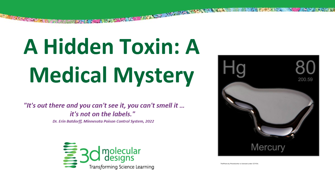
Aquaporin Mighty Model© A Hidden Toxin
-
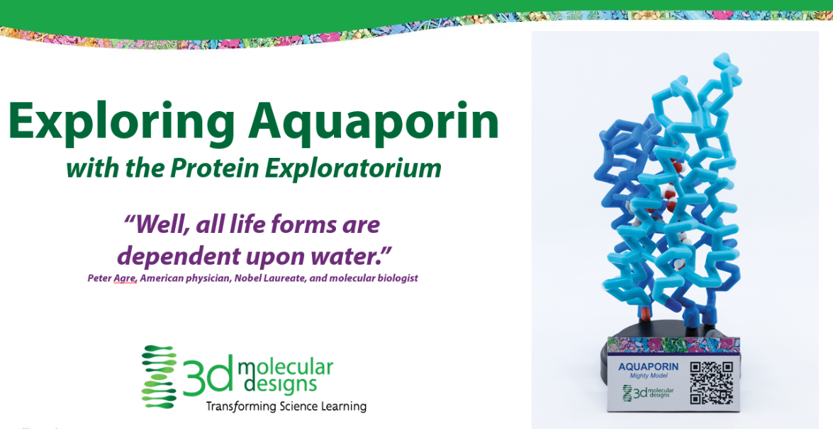
Exploring Aquaporin Mighty Model© with the Protein Exploratorium
-
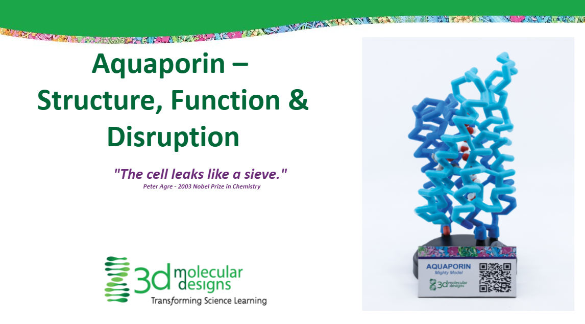
Aquaporin Mighty Model© Structure, Function & Distruption
-
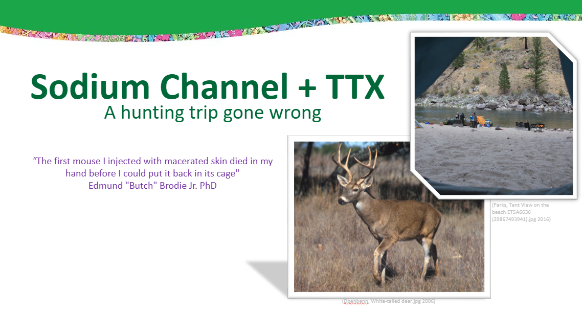
Sodium Channel Mighty Model©: A Hunting Trip Gone Wrong
-
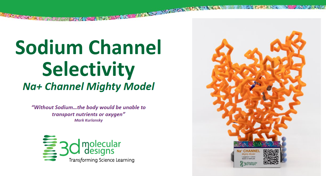
Sodium Channel Mighty Model© Selectivity
-
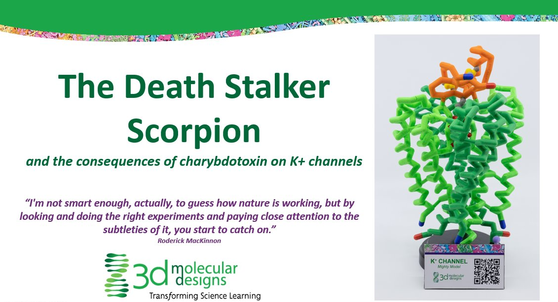
K+ Channel Mighty Model© The Death Stalker Scorpion
-
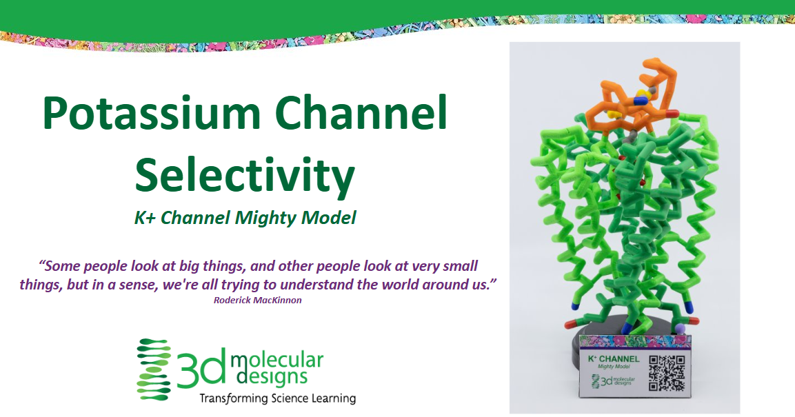
Potassium Channel Selectivity
























.jpg)