
Skip to content
header-top-2

◀ Back to Digital Modeling Hub
https://3dmoleculardesigns.com/hubfs/Codon%20Guides%20-%20Activities/Codon%20chart%20thumbnail.jpg
https://3dmoleculardesigns.com/hubfs/Digital%20Modeling%20Hub/Slide%20Screenshots/organ%20systems%20at%20work.jpg
https://3dmoleculardesigns.com/hubfs/Digital%20Modeling%20Hub/Slide%20Screenshots/building%20perfect%20cell%20screenshot.jpg
https://3dmoleculardesigns.com/hubfs/Digital%20Modeling%20Hub/Slide%20Screenshots/Antibody%20Production%20and%20Cellular%20Processes%20Screenshot.jpg
https://3dmoleculardesigns.com/hubfs/Digital%20Modeling%20Hub/Slide%20Screenshots/hemoglobinMM_screenshot.jpg
https://3dmoleculardesigns.com/hubfs/MOL%20Poster.jpg
https://3dmoleculardesigns.com/hubfs/Digital%20Modeling%20Hub/Slide%20Screenshots/6th%20grade%20activity%20.jpg
https://3dmoleculardesigns.com/hubfs/Digital%20Modeling%20Hub/Slide%20Screenshots/9th%20grade%20activity.jpg
https://3dmoleculardesigns.com/hubfs/Digital%20Modeling%20Hub/Slide%20Screenshots/MOLMK_slidedeck%20screenshot.jpg
https://3dmoleculardesigns.com/hubfs/Digital%20Modeling%20Hub/Slide%20Screenshots/MOL%20DMH%20pic.jpg
https://3dmoleculardesigns.com/hubfs/Digital%20Modeling%20Hub/Slide%20Screenshots/Stabilizing%20structures%20photo.jpg
https://3dmoleculardesigns.com/hubfs/Digital%20Modeling%20Hub/Slide%20Screenshots/protein%20tertiary%20structure%20picture.jpg
https://3dmoleculardesigns.com/hubfs/Digital%20Modeling%20Hub/Slide%20Screenshots/phage%20infection%20producrt%20image.jpg
https://3dmoleculardesigns.com/hubfs/Digital%20Modeling%20Hub/Slide%20Screenshots/phage%20replication%20product%20image.jpg
https://3dmd.3dmoleculardesigns.com/hubfs/Exploratorium%20Screenshots/AQP_Screenshot.png

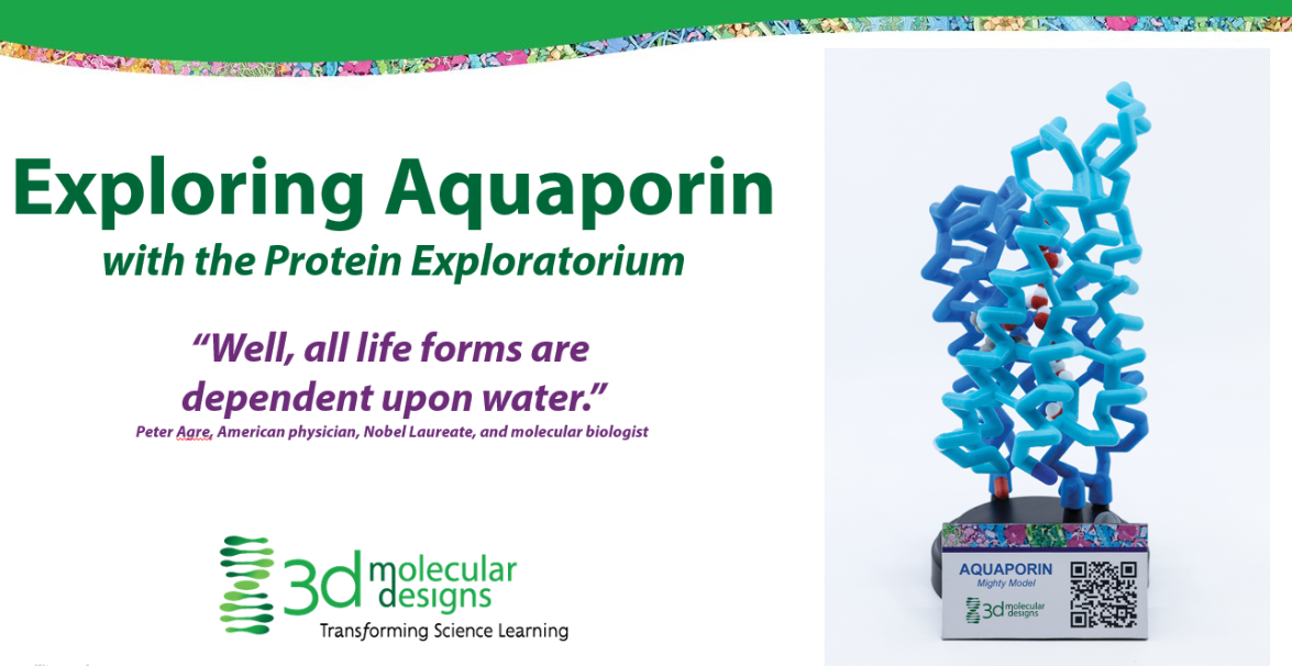
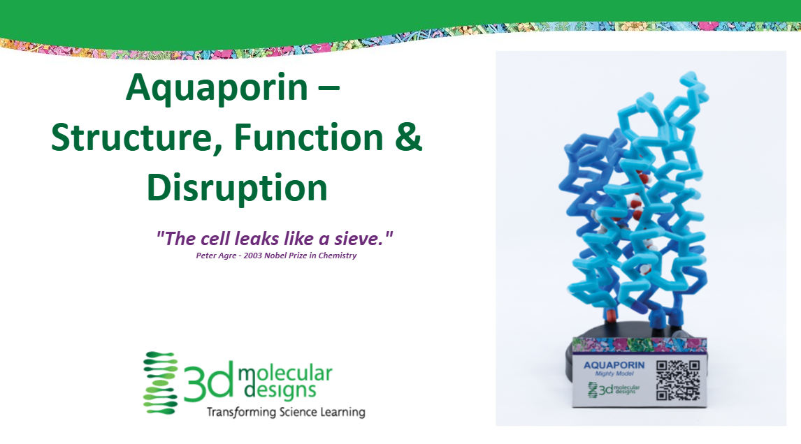
A 3D Molecular Design model of Aquaporin

https://3dmd.3dmoleculardesigns.com/hubfs/TeacherStudentGuideScreenshots/FlowofGeneticInformationKitTranslationStudentWorksheet_1.png

Student handouts for use with the purple translation mat in the Flow of Genetic Information Kit ©

https://3dmd.3dmoleculardesigns.com/hubfs/TeacherStudentGuideScreenshots/FlowofGeneticInformationKitTranslationStudentWorksheet_2.png
https://3dmd.3dmoleculardesigns.com/hubfs/TeacherStudentGuideScreenshots/PhospholipidAndMembraneTransportKItResourcesStudentHandout_1.png

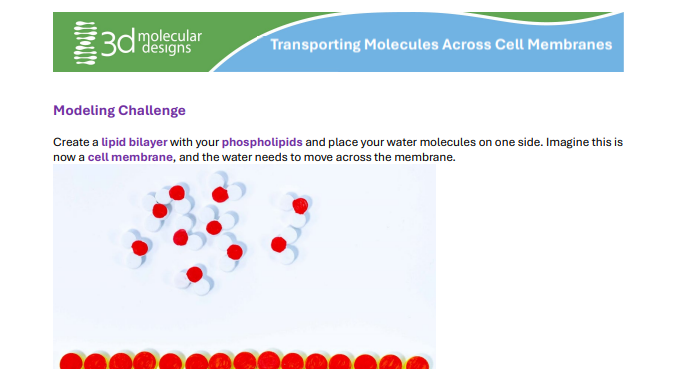
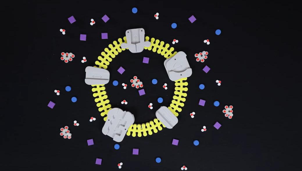
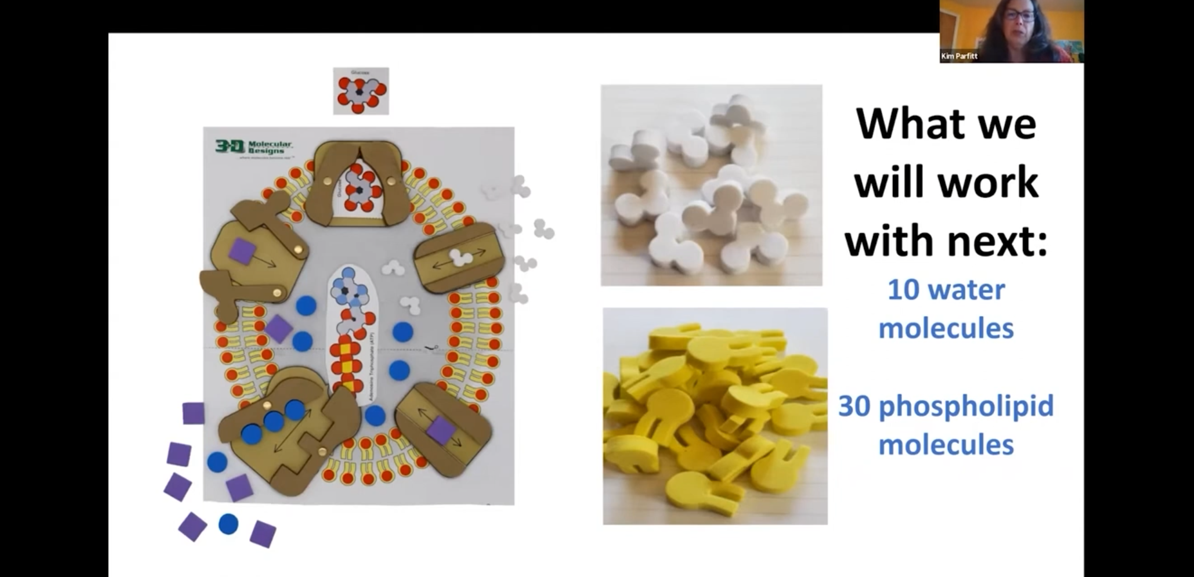
Students can model different conditions and then predict which way water will move, making vocabulary like hypertonic, hypotonic, and isotonic more memorable. This is a kit you will use over and over as your curriculum progresses to deepen student understanding.

https://3dmd.3dmoleculardesigns.com/hubfs/TeacherStudentGuideScreenshots/PhospholipidAndMembraneTransportKItResourcesTeacherHandout_1.png
https://3dmd.3dmoleculardesigns.com/hubfs/ArticulateScreenshots/DNA_MtP_1.png

A 3D Molecular Design Interactable about Monomers and Polymers

https://3dmd.3dmoleculardesigns.com/hubfs/ArticulateScreenshots/DNA_MtP_2.png
https://3dmd.3dmoleculardesigns.com/hubfs/VideoScreenshots/ComparingPhospholipidMonolayersMicellesAndBilayers_1.png

A 3D Molecular Design short content video about monolayers, micelles, and bilayers.

https://3dmd.3dmoleculardesigns.com/hubfs/VideoScreenshots/ComparingPhospholipidMonolayersMicellesAndBilayers_3.png
https://3dmd.3dmoleculardesigns.com/hubfs/Digital%20Modeling%20Hub/ArticulateScreenshots/waterMonomerToPolymer_1.jpg
https://3dmd.3dmoleculardesigns.com/hubfs/Digital%20Modeling%20Hub/ArticulateScreenshots/waterMonomerToPolymer_2.jpg
https://3dmd.3dmoleculardesigns.com/hubfs/AugmentedRealityScreenshots/SideChainExplorerAR_3.png

AR enhancements of the Amino Acid Starter Kit allow for the exploration of how sidechain properties drive protein folding

https://3dmd.3dmoleculardesigns.com/hubfs/AugmentedRealityScreenshots/SideChainExplorerAR_1.png
https://3dmd.3dmoleculardesigns.com/hubfs/AugmentedRealityScreenshots/SideChainExplorerAR_2.png
https://3dmoleculardesigns.com/hubfs/Digital%20Modeling%20Hub/Slide%20Screenshots/Screenshot_21-3-2025_11827_3dmd.sharepoint.com.jpeg
https://3dmoleculardesigns.com/hubfs/Digital%20Modeling%20Hub/Slide%20Screenshots/Screenshot_21-3-2025_14251_3dmd.sharepoint.com.jpeg
https://3dmd.3dmoleculardesigns.com/hubfs/ArticulateScreenshots/Sidechain_2_1.png

Part 1 of 3D Molecular Designs Side Chain Interactable

https://3dmd.3dmoleculardesigns.com/hubfs/ArticulateScreenshots/Sidechain_2_2.png
https://3dmd.3dmoleculardesigns.com/hubfs/VideoScreenshots/ComparingPurinesAndPyrimidines_1.png

A 3D Molecular Design short content video nvestigating purines and pyrimidines

https://3dmd.3dmoleculardesigns.com/hubfs/VideoScreenshots/ComparingPurinesAndPyrimidines_3.png
https://3dmoleculardesigns.com/hubfs/Digital%20Modeling%20Hub/Digital%20Modeling%20Hub%20Images/Screenshot%202025-01-22%20154627.png
https://3dmd.3dmoleculardesigns.com/hubfs/TeacherStudentGuideScreenshots/WaterKitResourcesTeacherGuide_1.png

Considered the gateway kit for students to develop model literacy, this is a great beginning-of-the-year activity.

https://3dmd.3dmoleculardesigns.com/hubfs/TeacherStudentGuideScreenshots/WaterKitResourcesStudentGuide_1.png
https://3dmoleculardesigns.com/hubfs/Digital%20Modeling%20Hub/Screenshot%202025-02-12%20131204.png
https://3dmd.3dmoleculardesigns.com/hubfs/TeacherStudentGuideScreenshots/ChromosomeConnectionsKitResourcesStudentHandout_1.png

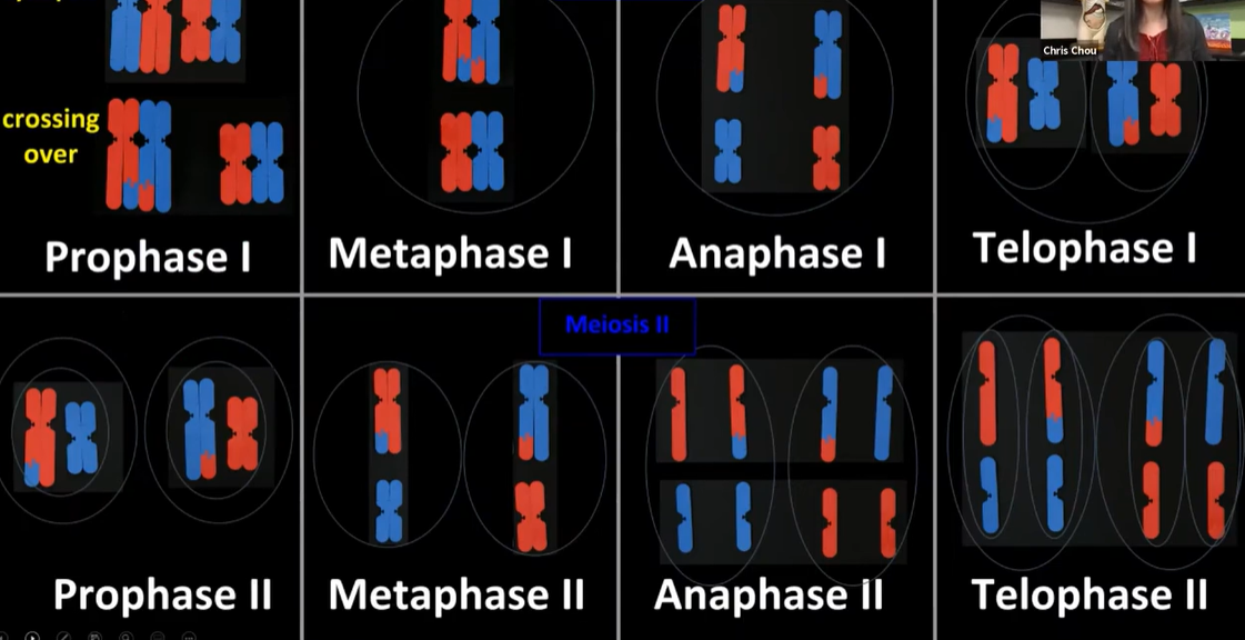
Conceptual understanding skyrockets as you invite students to investigate chromosomes at the microscopic and molecular scales! You will love the versatility of this kit as you explore the cell cycle, cell division, and genetics.

https://3dmd.3dmoleculardesigns.com/hubfs/TeacherStudentGuideScreenshots/ChromosomeConnectionsKitResourcesTeacherHandout_1.png
https://3dmd.3dmoleculardesigns.com/hubfs/TeacherStudentGuideScreenshots/BiotechnologyKitResourcesStudentHandout_1.png
  • Simulate PCR using flanking primers result to copy sequences of interest
  • Investigate how restriction endonucleases are used in DNA cloning
  • Explore the Sanger DNA sequencing method using chain-terminating fluorescent dideoxy nucleotide analogs
https://3dmd.3dmoleculardesigns.com/hubfs/TeacherStudentGuideScreenshots/BiotechnologyKitResourcesTeacherHandout_1.png
https://3dmd.3dmoleculardesigns.com/hubfs/Digital%20Modeling%20Hub/VideoScreenshots/DMH_Video_Intro_States%20of%20Matter.jpg

A 3D Molecular Design short content video about modeling the states of matter

https://3dmd.3dmoleculardesigns.com/hubfs/Digital%20Modeling%20Hub/VideoScreenshots/Screenshot%20states.png
https://3dmd.3dmoleculardesigns.com/hubfs/Digital%20Modeling%20Hub/VideoScreenshots/Screenshot%20states2.png
https://3dmd.3dmoleculardesigns.com/hubfs/TeacherStudentGuideScreenshots/AminoAcidStarterKitResourcesTeacherGuide_1.png

As your students begin to realize that DNA is code for an amino acid sequence that ultimately determines protein structure, their confidence soars!

https://3dmd.3dmoleculardesigns.com/hubfs/TeacherStudentGuideScreenshots/AminoAcidStarterKitResourcesStudentGuide_1.png
https://3dmoleculardesigns.com/hubfs/Digital%20Modeling%20Hub/Digital%20Modeling%20Hub%20Images/Screenshot%202025-02-11%20103845.png
https://3dmoleculardesigns.com/hubfs/Digital%20Modeling%20Hub/Digital%20Modeling%20Hub%20Images/Screenshot%202025-02-11%20103824.png
https://3dmoleculardesigns.com/hubfs/Digital%20Modeling%20Hub/Digital%20Modeling%20Hub%20Images/Screenshot%202025-02-11%20103916.png
https://3dmd.3dmoleculardesigns.com/hubfs/Digital%20Modeling%20Hub/VideoScreenshots/DMH_Video_Intro_Building%20a%20Phospholipid.jpg

A 3D Molecular Design short content video about the structure of phospholipids

https://3dmd.3dmoleculardesigns.com/hubfs/Digital%20Modeling%20Hub/VideoScreenshots/Screenshot%20building%20phospholipid2.png
https://3dmd.3dmoleculardesigns.com/hubfs/Digital%20Modeling%20Hub/VideoScreenshots/Screenshot%20building2.png
https://3dmd.3dmoleculardesigns.com/hubfs/Exploratorium%20Screenshots/ZincFingerExploritorium.png
A Jmol exploration of the 1ZAA Zinc Finger motif, complete with the amino acid sequence.
https://3dmd.3dmoleculardesigns.com/hubfs/AnabolicScreenshot.png

3D Molecular Designs Anabolic Interactable

https://3dmd.3dmoleculardesigns.com/hubfs/Digital%20Modeling%20Hub/Digital%20Modeling%20Hub%20Images/membraneMonomerToPolymer_1%201.jpg
https://3dmd.3dmoleculardesigns.com/hubfs/Digital%20Modeling%20Hub/Digital%20Modeling%20Hub%20Images/membraneMonomerToPolymer_2%201.jpg
https://3dmd.3dmoleculardesigns.com/hubfs/TeacherStudentGuideScreenshots/ChromosomeConnectionsKitResourcesStudentHandout_1.png

Conceptual understanding skyrockets as you invite students to investigate chromosomes at the microscopic and molecular scales! You will love the versatility of this kit as you explore the cell cycle, cell division, and genetics.

https://3dmd.3dmoleculardesigns.com/hubfs/TeacherStudentGuideScreenshots/ChromosomeConnectionsKitResourcesTeacherHandout_1.png
https://3dmoleculardesigns.com/hubfs/Screenshot%202025-02-26%20123302.png

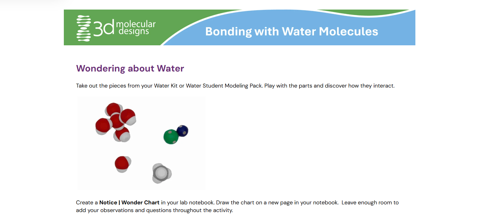
In this introductory lesson, students explore hydrogen and covalent bonds within and between molecules. Working through this independent activity with their physical models, lab notebook, and several different molecular representations of water, they learn about polarity and will model cohesion and hydrogen bonding.

https://3dmd.3dmoleculardesigns.com/hubfs/ArticulateScreenshots/BindingChem_1.png

A 3D Molecular Design Interactable about Binding Chemicals

https://3dmd.3dmoleculardesigns.com/hubfs/ArticulateScreenshots/BindingChem_2.png
https://3dmd.3dmoleculardesigns.com/hubfs/AugmentedRealityScreenshots/TransferringModelsWithTheFlowOfGeneticInformationKitAR_1.png

AR enhancements of DNA show the similarities and differences between multiple DNA representations

https://3dmd.3dmoleculardesigns.com/hubfs/AugmentedRealityScreenshots/ProteinTranlsationWithTheFlowOfGeneticInformationKitAR_2.png
https://3dmd.3dmoleculardesigns.com/hubfs/ArticulateScreenshots/BindingShape_1.png

A 3D Molecular Design Interactable about Shapes and their binding properties

https://3dmd.3dmoleculardesigns.com/hubfs/ArticulateScreenshots/BindingShape_2.png
https://3dmoleculardesigns.com/hubfs/Digital%20Modeling%20Hub/Digital%20Modeling%20Hub%20Images/Screenshot%202025-02-26%20130911.png
https://3dmd.3dmoleculardesigns.com/hubfs/TeacherStudentGuideScreenshots/GlobinFoldingKitTeacherHandout_1.png

Fun, self-correcting, and intuitive, this kit results in confidence and learning that lasts for years.

https://3dmd.3dmoleculardesigns.com/hubfs/TeacherStudentGuideScreenshots/GlobinFoldingKitStudentHandout_1.png
https://3dmoleculardesigns.com/hubfs/Screenshot%202025-01-27%20102739.png
https://learn.3dmoleculardesigns.com/hubfs/IMG_0012.jpg

As your students begin to realize that DNA is code for an amino acid sequence that ultimately determines protein structure, their confidence soars!

https://3dmd.3dmoleculardesigns.com/hubfs/AugmentedRealityScreenshots/TransferringModelsWithTheDynamicDNAKitAR_3.png

AR enhancements of DNA show the similarities and differences between multiple DNA representations

https://3dmd.3dmoleculardesigns.com/hubfs/AugmentedRealityScreenshots/TransferringModelsWithTheDynamicDNAKitAR_2.png
https://3dmd.3dmoleculardesigns.com/hubfs/AugmentedRealityScreenshots/TransferringModelsWithTheDynamicDNAKitAR_1.png
https://3dmd.3dmoleculardesigns.com/hubfs/Screenshot%202024-10-17%20115002.png

Students will create a model of a protein based on the 3D coordinates displayed on a webpage.

https://3dmd.3dmoleculardesigns.com/hubfs/Screenshot%202024-10-17%20115020.png

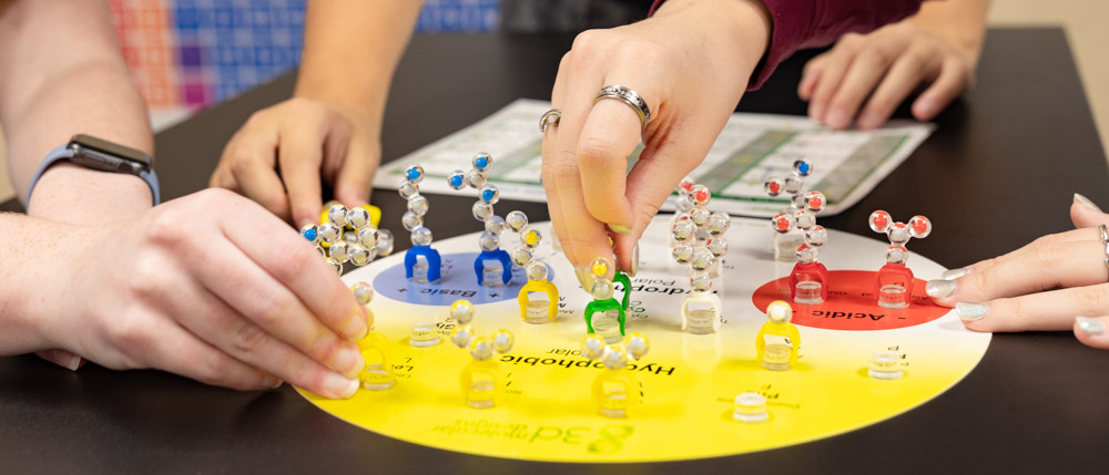
This hands-on modeling kit is designed to help students visualize protein structures and how a protein's unique shape impacts its function.

https://3dmd.3dmoleculardesigns.com/hubfs/Screenshot%202024-10-17%20115033.png

Students will explore protein structures on web pages to identify similarities and differences.

https://3dmd.3dmoleculardesigns.com/hubfs/VideoScreenshots/ExploringSoubilityofAlchohols1.png

A 3D Molecular Design video about the Solubility of Alcohols

https://3dmd.3dmoleculardesigns.com/hubfs/VideoScreenshots/ExploringSolubilityOfAlcohols_3.png
https://3dmd.3dmoleculardesigns.com/hubfs/VideoScreenshots/BuildingATriglyceride_1.png

A 3D Molecular Design short content video about triglyceride structure, oils, and fats

https://3dmd.3dmoleculardesigns.com/hubfs/VideoScreenshots/BuildingATriglyceride_3.png
https://learn.3dmoleculardesigns.com/hubfs/Digital%20Modeling%20Hub%20Images/jmolTrainingGuide.jpg

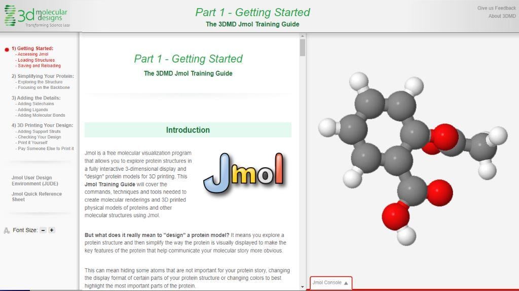
The 3DMD Jmol Training Guide covers how to access Jmol, design a protein model, and export it for 3D printing.

https://3dmoleculardesigns.com/hubfs/Screenshot%202025-01-24%20093258.png
https://3dmoleculardesigns.com/hubfs/Digital%20Modeling%20Hub/Screenshot%202025-02-26%20150749.png
https://3dmoleculardesigns.com/hubfs/Digital%20Modeling%20Hub/Digital%20Modeling%20Hub%20Images/MakingtheCut%20CRISPR%20product%20image.jpg

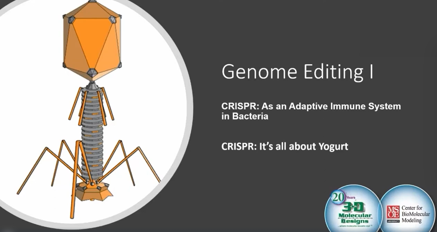
Set your students up for successful CRISPR investigations by first exploring how bacteria seek and destroy viral invaders.

https://3dmoleculardesigns.com/hubfs/Digital%20Modeling%20Hub/Slide%20Screenshots/Adaptive%20Immunity%20pic.jpg

Set your students up for successful CRISPR investigations by first exploring how bacteria seek and destroy viral invaders.

https://3dmd.3dmoleculardesigns.com/hubfs/Screenshot%202024-10-16%20114837.png

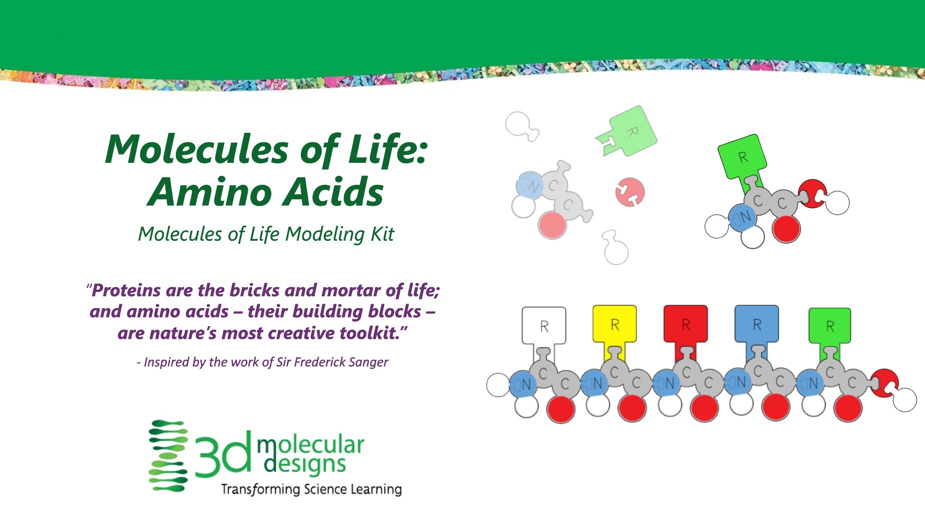
Students explore the properties of amino acids and how they are connected to form a polypeptide.

https://3dmd.3dmoleculardesigns.com/hubfs/Screenshot%202024-10-16%20114846.png

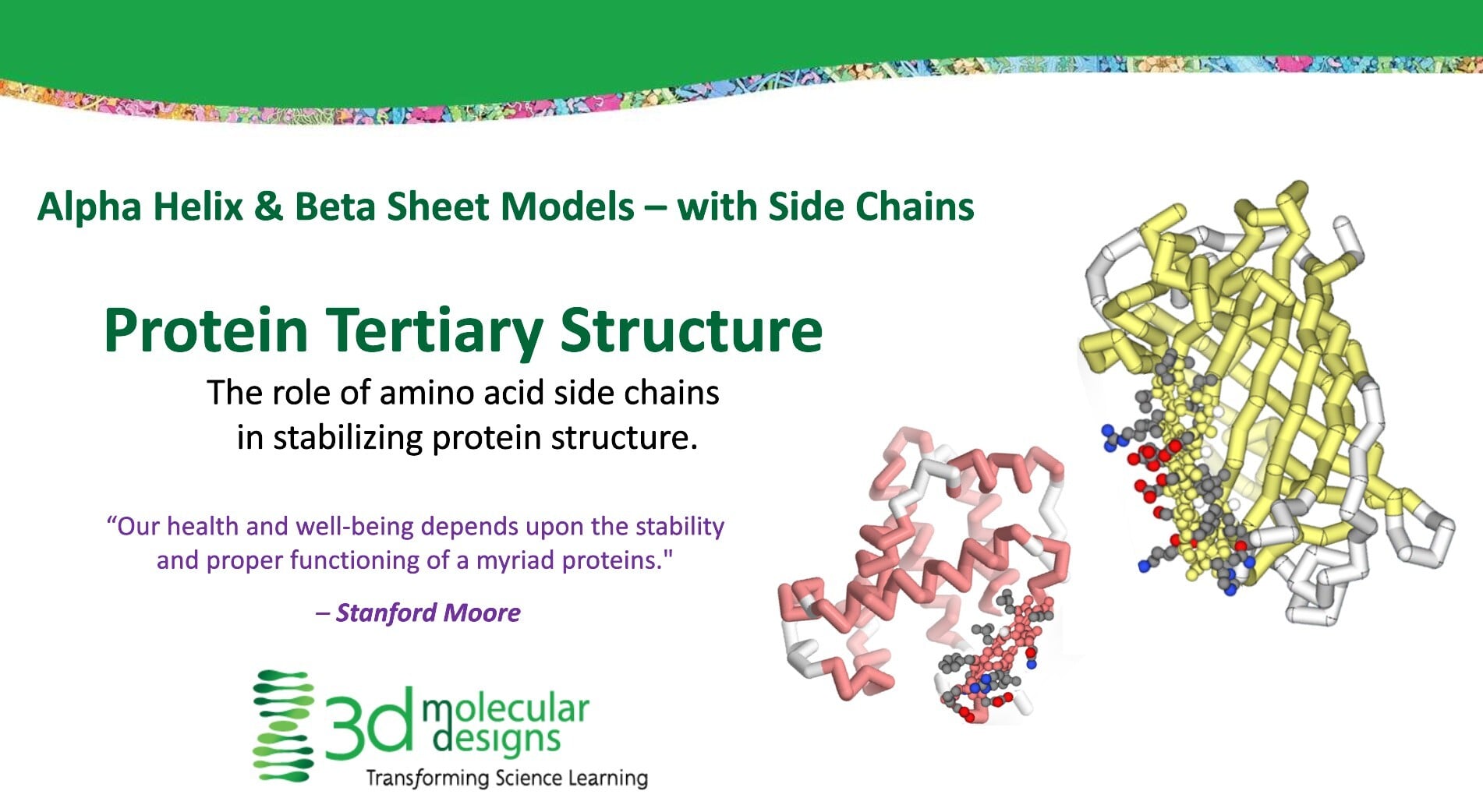
Students will learn about alpha helices and beta sheets and how they are displayed in molecular visualization software.

https://3dmd.3dmoleculardesigns.com/hubfs/Screenshot%202024-10-16%20114901.png

Students will explore how the properties of the amino acid sidechains influence the tertiary structure of a protein.

https://3dmd.3dmoleculardesigns.com/hubfs/AugmentedRealityScreenshots/ExploringStatesofMatterAR_2.png

AR enhancements of the Water kit allow students to explore and model the states of matter

https://3dmd.3dmoleculardesigns.com/hubfs/AugmentedRealityScreenshots/ExploringStatesofMatterAR_1.png
https://3dmd.3dmoleculardesigns.com/hubfs/VideoScreenshots/ComponentsOfAPhospholipid_1.png

A 3D Molecular Design short content video about the diversity of phospholipids

https://3dmd.3dmoleculardesigns.com/hubfs/VideoScreenshots/ComponentsOfAPhospholipid_2.png
https://3dmd.3dmoleculardesigns.com/hubfs/Digital%20Modeling%20Hub/TeacherStudentGuideScreenshots/NeuromuscularSynapsePosterColoringPage.png
https://3dmd.3dmoleculardesigns.com/hubfs/ArticulateScreenshots/Sidechain_1_1.png

Part 2 of 3D Molecular Designs Side Chain Interactable

https://3dmd.3dmoleculardesigns.com/hubfs/ArticulateScreenshots/Sidechain_1_2.png
https://3dmd.3dmoleculardesigns.com/hubfs/ArticulateScreenshots/Catabolic_1.png

A 3D Molecular Design Interactable about Catabolic reactions

https://3dmd.3dmoleculardesigns.com/hubfs/ArticulateScreenshots/Catabolic_2.png
https://3dmd.3dmoleculardesigns.com/hubfs/Exploratorium%20Screenshots/ARTrainingApp.png
Free resources to use with your class as they begin to explore our AR app, available in both the Apple and Google Play stores.
https://3dmd.3dmoleculardesigns.com/hubfs/TeacherStudentGuideScreenshots/MakingTheCutWithCRISPERCas9StudentHandout_1.png

Tim Herman, PhD, 3D Molecular Designs' founder and scientific officer, explores the CRISPR/Cas9 system.

https://3dmd.3dmoleculardesigns.com/hubfs/TeacherStudentGuideScreenshots/MakingTheCutWithCRISPERCas9StudentHandout_2.png
https://3dmd.3dmoleculardesigns.com/hubfs/AugmentedRealityScreenshots/ProteinTranlsationWithTheFlowOfGeneticInformationKitAR_2.png

AR enhancements of the Flow of Genetic Information Kit allow students to model protein synthesis and investigate how different antibiotics inhibit protein production

https://3dmd.3dmoleculardesigns.com/hubfs/AugmentedRealityScreenshots/ProteinTranlsationWithTheFlowOfGeneticInformationKitAR_1.png
https://3dmd.3dmoleculardesigns.com/hubfs/TeacherStudentGuideScreenshots/FlowofGeneticInformationKitResourcesStudentGuide_1.png

A 3D Molecular Design video demonstrating the Flow of Genetic Information kit

https://3dmd.3dmoleculardesigns.com/hubfs/TeacherStudentGuideScreenshots/FlowofGeneticInformationKitResourcesTeacherGuide_1.png
https://3dmd.3dmoleculardesigns.com/hubfs/AugmentedRealityScreenshots/DiscoveringPolarityAR_1.png

AR enhancements of the Water Kit allow students to discover the molecular reason for water polarity

https://3dmd.3dmoleculardesigns.com/hubfs/AugmentedRealityScreenshots/DiscoveringPolarityAR_2.png
https://3dmd.3dmoleculardesigns.com/hubfs/AugmentedRealityScreenshots/FoldingRealProteinsZincFingersAR_2.png

AR enhancements of the Amino Acid Starter Kit allow students to begin investigating the different levels of protein folding

https://3dmd.3dmoleculardesigns.com/hubfs/AugmentedRealityScreenshots/FoldingRealProteinsZincFingersAR_1.png
https://3dmoleculardesigns.com/hubfs/Digital%20Modeling%20Hub/Digital%20Modeling%20Hub%20Images/Screenshot%202025-02-04%20115310.png
https://3dmd.3dmoleculardesigns.com/hubfs/7P0A6231.webp

Dr. Goodsell creates cellular landscapes that accurately illustrate the size, shape, and distribution of proteins in a B-cell

https://3dmd.3dmoleculardesigns.com/hubfs/Exploratorium%20Screenshots/MoleculeMakerImage.png

Load an existing SMILE file or build your own small molecules. Export them as images for use in a document or presentation or export them as .STL files for 3D Printing

https://3dmoleculardesigns.com/hubfs/Digital%20Modeling%20Hub/Digital%20Modeling%20Hub%20Images/physical%20chemical%20props%20of%20water.jpg

Considered the gateway kit for students to develop model literacy, this is a great beginning-of-the-year activity.

https://3dmd.3dmoleculardesigns.com/hubfs/AugmentedRealityScreenshots/ExtendingTheDNADoubleHelixWithDynamicDNAKitAR_3.png

AR enhancements of the Dynamic DNA kit allow students to begin investigating DNA - protein interactions

https://3dmd.3dmoleculardesigns.com/hubfs/AugmentedRealityScreenshots/ExtendingTheDNADoubleHelixWithDynamicDNAKitAR_2.png
https://3dmd.3dmoleculardesigns.com/hubfs/AugmentedRealityScreenshots/ExtendingTheDNADoubleHelixWithDynamicDNAKitAR_1.png
https://3dmoleculardesigns.com/hubfs/Digital%20Modeling%20Hub/Digital%20Modeling%20Hub%20Images/Screenshot%202025-01-23%20143429.png

Webinar Series:

CRISPR Cas-9 - A Powerful New Tool for Editing the Human Genome

CRISPR Innovations: Clever and Cleverer

Tim Herman, PhD - 3D Molecular Designs

https://3dmd.3dmoleculardesigns.com/hubfs/TeacherStudentGuideScreenshots/EnzymesInActionKitResourcesStudent_1.png

Excellent at revealing student thinking, this kit can complement traditional classroom assignments like the catalase or amylase labs,

https://3dmd.3dmoleculardesigns.com/hubfs/TeacherStudentGuideScreenshots/EnzymesInActionKitResourcesStudent_2.png
https://3dmoleculardesigns.com/hubfs/Digital%20Modeling%20Hub/Digital%20Modeling%20Hub%20Images/Screenshot%202025-02-07%20133722.png

Tim Herman, PhD, 3D Molecular Designs' founder and scientific officer, explores the CRISPR/Cas9 system.

https://3dmd.3dmoleculardesigns.com/hubfs/TeacherStudentGuideScreenshots/WaterKitResourcesTeacherGuide_1.png

Considered the gateway kit for students to develop model literacy, this is a great beginning-of-the-year activity.

https://3dmd.3dmoleculardesigns.com/hubfs/TeacherStudentGuideScreenshots/WaterKitResourcesStudentGuide_1.png
https://3dmoleculardesigns.com/hubfs/Digital%20Modeling%20Hub/Screenshot%202025-01-22%20093858.png
https://3dmoleculardesigns.com/hubfs/Digital%20Modeling%20Hub/Digital%20Modeling%20Hub%20Videos/PMTK_parfitt.png
https://3dmd.3dmoleculardesigns.com/hubfs/TeacherStudentGuideScreenshots/InsulinmRNAToProteinKitStudentHandout_1.png

Your students can connect protein synthesis to blood sugar regulation as they fold their own insulin model.

https://3dmd.3dmoleculardesigns.com/hubfs/TeacherStudentGuideScreenshots/InsulinmRNAToProteinKitStudentHandout_2.png
https://3dmoleculardesigns.com/hubfs/Digital%20Modeling%20Hub/Digital%20Modeling%20Hub%20Images/Screenshot%202025-02-04%20150233.png
https://3dmoleculardesigns.com/hubfs/Digital%20Modeling%20Hub/Digital%20Modeling%20Hub%20Images/Screenshot%202025-02-06%20142527.png
https://3dmd.3dmoleculardesigns.com/hubfs/TeacherStudentGuideScreenshots/NaCLLatticeStudentHandout_1.png

Keep this mesmerizing model on your desk to invite student questions


https://3dmd.3dmoleculardesigns.com/hubfs/TeacherStudentGuideScreenshots/NaCLLatticeTeacherHandout_1.png
https://3dmd.3dmoleculardesigns.com/hubfs/TeacherStudentGuideScreenshots/DNAStarterKitResourcesTeacherHandout_1.png

This easy-to-build model is a perfect starting point that students can use to test their ideas and transfer their understanding to other, more complex models.


https://3dmd.3dmoleculardesigns.com/hubfs/TeacherStudentGuideScreenshots/DNAStarterKitResourcesTeacherHandout_2.png
https://3dmd.3dmoleculardesigns.com/hubfs/Digital%20Modeling%20Hub/VideoScreenshots/DMH_Video_Intro_RNA%20Processing%20(1).jpg

A 3D Molecular Design short content video about RNA processing, including polyadenylation, methyl caps, and more.

https://shop.3dmoleculardesigns.com/cdn/shop/files/INSFK_A_S4_Web.jpg

Your students can connect protein synthesis to blood sugar regulation as they fold their own insulin model.

https://3dmd.3dmoleculardesigns.com/hubfs/Digital%20Modeling%20Hub/VideoScreenshots/DMH_Video%20Intro_Chromosome%20and%20Gene%20Structures.jpg

A 3D Molecular Design short content video about chromosome structure.

https://3dmd.3dmoleculardesigns.com/hubfs/Exploratorium%20Screenshots/JUDEImage.png

A molecular structure file includes the X, Y, and Z coordinates to position each atom in a molecule in 3D space. The buttons below will load structure files in the Jmol window to the right.

https://shop.3dmoleculardesigns.com/cdn/shop/files/BTK_Seq_glamor2.jpg

Amplify understanding as students model the molecular processes in your pGLO or crime scene labs.

https://3dmd.3dmoleculardesigns.com/hubfs/Exploratorium%20Screenshots/ProteinExploritorium_1.png
A Jmol exploration of any PDB file you choose, complete with amino acid sequence.
https://3dmd.3dmoleculardesigns.com/hubfs/TeacherStudentGuideScreenshots/DynamicDNAKitResourcesStudentGuide_1.png

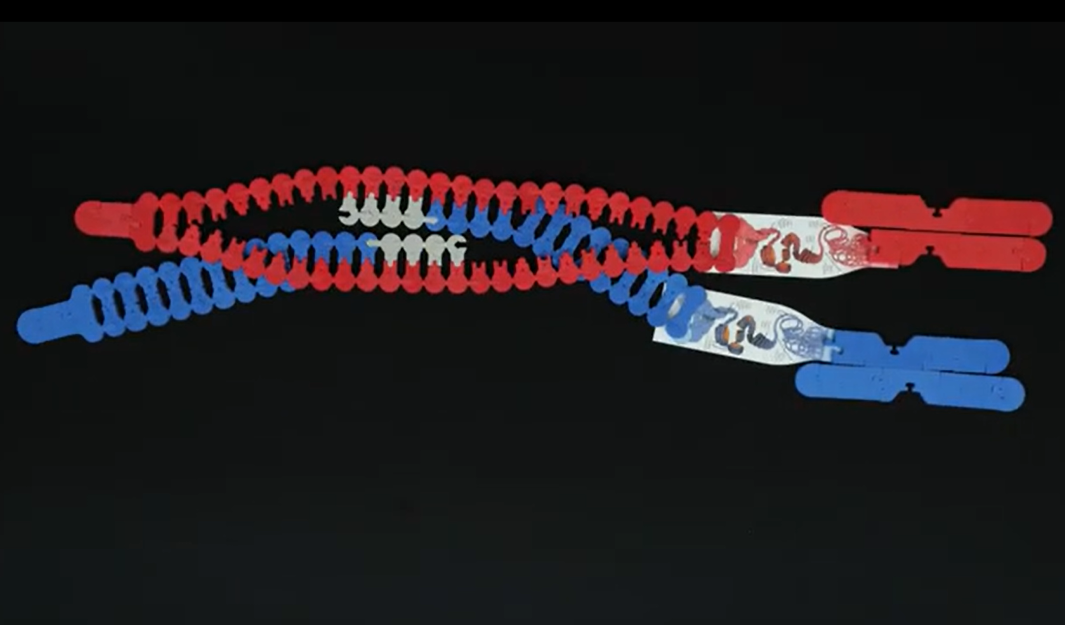
Thanks to our patented design innovations, the structure of this molecule is revealed to students through their own investigation.

https://3dmd.3dmoleculardesigns.com/hubfs/TeacherStudentGuideScreenshots/DynamicDNAKitResourcesTeacherGuide_1.png
https://3dmd.3dmoleculardesigns.com/hubfs/TeacherStudentGuideScreenshots/TourOfAHumanCellTeacherGuides_1.png

Dr. Goodsell creates cellular landscapes that accurately illustrate the size, shape, and distribution of proteins in a B-cell

https://3dmd.3dmoleculardesigns.com/hubfs/TeacherStudentGuideScreenshots/TourOfAHumanCellTeacherGuides_2.png
https://3dmd.3dmoleculardesigns.com/hubfs/video16Cover.jpg

Learn how to prepare your Modeling Tie to create an accurate model of a protein.

https://3dmd.3dmoleculardesigns.com/hubfs/video15Cover.jpg

Learn how to fold your Modeling Tie into an accurate model of a protein.

https://3dmd.3dmoleculardesigns.com/hubfs/Digital%20Modeling%20Hub/VideoScreenshots/DMH_Video_Intro_ID%20Regions%20Chromosome.jpg

A 3D Molecular Design short content video about p arms, q arms, telomeres and more

https://3dmd.3dmoleculardesigns.com/hubfs/Digital%20Modeling%20Hub/VideoScreenshots/Screenshot%20reg%20chromosome.png
https://3dmd.3dmoleculardesigns.com/hubfs/Digital%20Modeling%20Hub/VideoScreenshots/Screenshot%20reg%20chromosome2.png
https://shop.3dmoleculardesigns.com/cdn/shop/files/SCK1_glamor.jpg

Your students will uncover how electrical and chemical signals allow us to move, breathe, and think, then predict what happens when things go wrong.

https://learn.3dmoleculardesigns.com/hubfs/Product%20Promo%20Photos/Chromosome%20Connections%20Kit/Chromosome%20Connections%20Kits%205.jpg

Conceptual understanding skyrockets as you invite students to investigate chromosomes at the microscopic and molecular scales! You will love the versatility of this kit as you explore the cell cycle, cell division, and genetics.

https://3dmd.3dmoleculardesigns.com/hubfs/TeacherStudentGuideScreenshots/NeuronModelingKitStudentHandout_1.png

Get your students' own neurons firing with these models.

https://3dmd.3dmoleculardesigns.com/hubfs/TeacherStudentGuideScreenshots/NeuronModelingKitTeacherHandout_1.png
https://learn.3dmoleculardesigns.com/hubfs/Lending%20Library/Dynamic%20DNA%20Kit/DDNA_LL_glamour3_Web.jpg

Thanks to our patented design innovations, the structure of this molecule is revealed to students through their own investigation.

https://3dmd.3dmoleculardesigns.com/hubfs/Digital%20Modeling%20Hub/VideoScreenshots/DMH_Video_Intro_Exploring%20Saturated.jpg

A 3D Molecular Design Short Content video modeling different types of solutions

https://3dmd.3dmoleculardesigns.com/hubfs/Digital%20Modeling%20Hub/VideoScreenshots/Screenshot%20unsat%20sat%20supersat%201.png
https://3dmd.3dmoleculardesigns.com/hubfs/Digital%20Modeling%20Hub/VideoScreenshots/sat%20super4.png
https://3dmoleculardesigns.com/hubfs/Digital%20Modeling%20Hub/Digital%20Modeling%20Hub%20Images/Screenshot%202025-01-22%20134931.png
https://3dmd.3dmoleculardesigns.com/hubfs/TeacherStudentGuideScreenshots/MapOfTheHumanGlobinGeneTeacherHandout_1.png

You will astonish your students with how much information is in one gene just by unrolling this 15-foot map in the classroom.

https://3dmd.3dmoleculardesigns.com/hubfs/TeacherStudentGuideScreenshots/MapOfTheHumanGlobinGeneStudentHandout_1.png
https://3dmd.3dmoleculardesigns.com/hubfs/Screenshot%202024-10-16%20155507.png

Cytochrome C is an important protein structure involved in carrying electrons during respiration.

https://3dmd.3dmoleculardesigns.com/hubfs/Screenshot%202024-10-16%20155452.png

Immunoglobulin Folds are an important protein motif found immunology proteins such as antibodies. 

https://3dmd.3dmoleculardesigns.com/hubfs/Screenshot%202024-10-16%20155500.png

Zinc Fingers are an important family of protein structures involved in regulating access to DNA in the nucleus of our cells. 

https://3dmd.3dmoleculardesigns.com/hubfs/TeacherStudentGuideScreenshots/SynapseContstructionKitStudentHandout_1.png

Puzzling promotes sensemaking when students engineer an enzyme's active site.

https://3dmd.3dmoleculardesigns.com/hubfs/TeacherStudentGuideScreenshots/SubstrateSpecificityKitStudentHandout_1.png
https://3dmd.3dmoleculardesigns.com/hubfs/TeacherStudentGuideScreenshots/SynapseContstructionKitTeacherHandout_1.png

Your students will uncover how electrical and chemical signals allow us to move, breathe, and think, then predict what happens when things go wrong.

https://3dmd.3dmoleculardesigns.com/hubfs/TeacherStudentGuideScreenshots/SynapseContstructionKitStudentHandout_1.png
https://3dmd.3dmoleculardesigns.com/hubfs/TeacherStudentGuideScreenshots/ModelingMiniTooberTeacherHandout_1.png
https://3dmd.3dmoleculardesigns.com/hubfs/Digital%20Modeling%20Hub/VideoScreenshots/coronavirus2.png
https://3dmd.3dmoleculardesigns.com/hubfs/Digital%20Modeling%20Hub/VideoScreenshots/coronavirus1.png
https://3dmd.3dmoleculardesigns.com/hubfs/Digital%20Modeling%20Hub/VideoScreenshots/Coronavirus%20slide.jpeg
https://3dmd.3dmoleculardesigns.com/hubfs/Digital%20Modeling%20Hub/TeacherStudentGuideScreenshots/DataDilemmaModelingTheScientificMethod.png
https://3dmd.3dmoleculardesigns.com/hubfs/Digital%20Modeling%20Hub/TeacherStudentGuideScreenshots/DataDilemmmaTeacherNotes.png
https://3dmoleculardesigns.com/hubfs/PSMP_A_P1_glamour_Web-1.jpg
https://3dmoleculardesigns.com/hubfs/DSMP_A_P1_glamour_Web-1.jpg
https://3dmoleculardesigns.com/hubfs/CSMP_A_P1_glamour_Web-1.jpg
https://3dmoleculardesigns.com/hubfs/MSMP_A_P1_glamour_Web-1.jpg
https://3dmoleculardesigns.com/hubfs/WSMP_A_P1_glamour_Web-1.jpg
https://3dmoleculardesigns.com/hubfs/NSMP_A_P1_glamour_Web-1.jpg

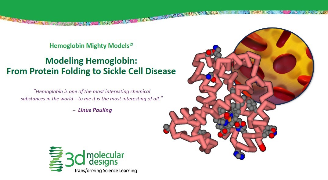
Modeling Hemoglobin: From Protein Folding to Sickle Cell Disease Slide Deck

Access: Available as a Free Resource
Link: See Student Handouts Below

Activity Type: Activity Slide Decks
Associated Product: Hemoglobin Mighty Model©


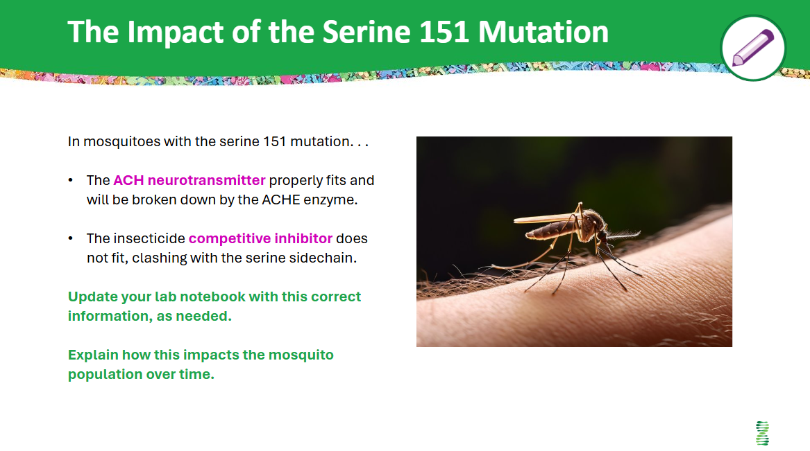
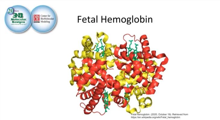
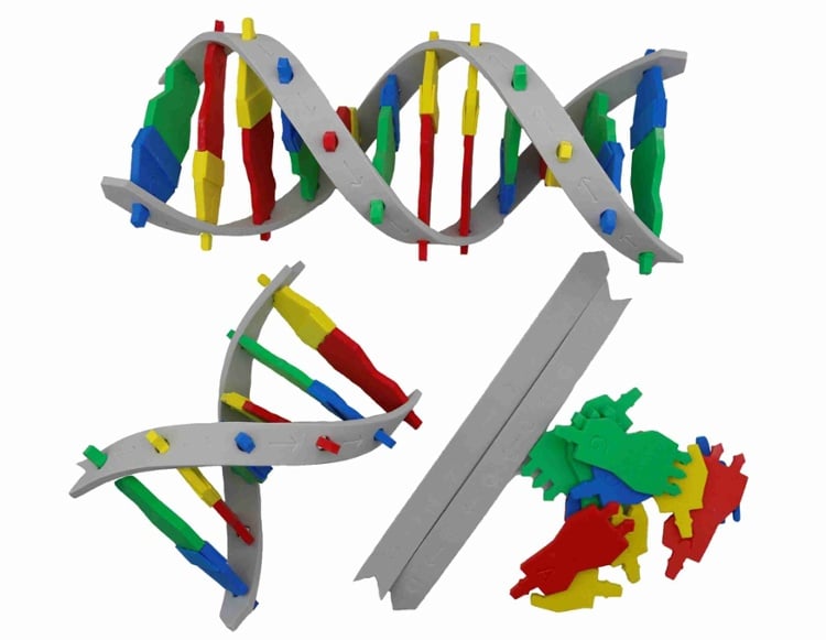
Students use 3D alpha- and beta-globin models to investigate all four levels of protein structure while connecting molecular details to a real patient story and the genetics of sickle cell disease. Anchored in the case of Victoria Gray, students begin at the organism and organ-system level (symptoms, circulation, red blood cell shape), then zoom in to the cellular and molecular scales to explore how hemoglobin’s structure enables oxygen transport—and how a single amino acid substitution (glutamic acid to valine at position six in beta-globin) can distort red blood cells and impair blood flow. Along the way, they trace the globin backbone from N- to C-terminus, link “bends” in the ribbon to individual amino acids, examine selected sidechains, and classify them as hydrophilic or hydrophobic to explain how polarity, charge, and the hydrophobic effect drive folding into tertiary and quaternary structures and position heme groups for oxygen binding.
Designed primarily for grades 9–12 Biology, Honors Biology, and AP Biology, this activity fits comfortably into one 60–75 minute class period for the core investigation, with an additional 45–60 minutes if you choose to use all extensions and assessments. It pairs especially well with 3DMD’s Amino Acid Starter Kit and Protein Student Modeling resources, giving students a concrete bridge from amino acid properties to full protein models. The slide deck includes student-facing prompts and rich teacher notes, with built-in routines such as Notice–Wonder charts, quiet thinking time, turn-and-talk discussion, digital and physical modeling, and frequent formative checks to surface misconceptions about protein structure, red blood cell function, and genetic disease.

For advanced high school or introductory college courses, optional extensions connect protein structure to gene structure and biotechnology. Students can map the 146–amino-acid beta-globin chain back to its gene with exons and introns, examine carbon monoxide as a competitive inhibitor at the heme site, and discuss how CRISPR-based genome editing has been used clinically to treat sickle cell disease, including in Victoria Gray’s case. These extensions support NGSS performance expectations HS-LS1-1 and HS-LS1-2 (structure and function of proteins), HS-LS3-1 and HS-LS3-2 (inheritance and variation of traits, including the Glu→Val mutation), and highlight crosscutting concepts of Structure and Function, Cause and Effect, and Systems and System Models, making the lesson a strong fit for units on molecular genetics, heredity, human physiology, and biotechnology.

Plan for one model per lab group – students can use either the Beta-Globin Mighty Model, or the Alpha-Globin Mighty Model. When the Sickle Cell mutation is discussed, lab groups can combine, or those with an alpha-globin model can engage with their models and the images in the slide deck. If you have 2 alpha- and 2 beta-globin models, have your students combine them to experience quaternary structure! Amino acid sidechain models are also needed – either from the Amino Acid Starter Kit or print free paper versions.

Slide Decks incorporate all the information an educator needs to run the activity in one file. Slides that are meant for student viewing feature a green top banner. Slides that are meant only for teacher viewing feature a purple top banner and are hidden in slide-show mode. Each student slide will include a prompting icon in the top right corner. These icons on each slide cue your students to the type of activity they will be doing. Student slides provide notes (teaching notes below the slide) with specific information to assist you in facilitating the activity using effective modeling and education practices.   

Become an Educator to get access to the answer keys!