





 What is a Protein?
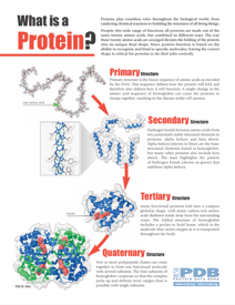
This two-page PDF introduces what proteins are, how their 3-dimensional structure relates to their function, and what jobs they perform in our cells.
Proteins are some of the most important molecules in biochemistry. They are long strands that fold up into an endless variety of 3-dimensional shapes and sizes, each uniquely structured to perform a specific function in the body.
Because proteins are such large and complex molecules, scientists often describe their shape using four layers of protein structure. Explore each of the four layers using the interactive webpages linked below.
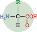
All proteins are made from small molecules called amino acids that are joined together like links in a chain. The sequence of amino acids in a protein determines its structure and shape.
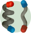
Protein chains fold into complex 3-dimensional shapes with two common secondary structures, alpha helices and beta sheets.

Proteins continue to fold into an overall 3-dimensional shape with precise arrangements of their secondary structures.
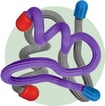
Some proteins comprise more than one amino acid chain, forming stable multi-chain assemblies.
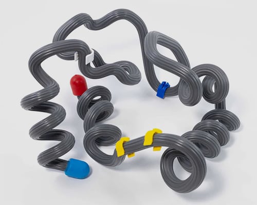 Protein Structure Modeling Kit
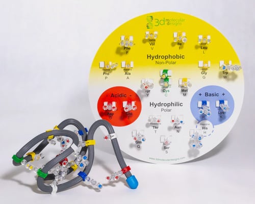
Learn about protein Structure and function with the hands-on kit that inspired the protein modeling event.
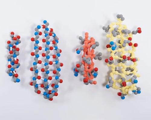 Alpha Helix & Beta Sheet Structure Sets
Explore how the repeating N-C-C pattern can result in two very different secondary structures and search for patterns based on the hydrophobic or hydrophilic properties of each backbone’s side chains.
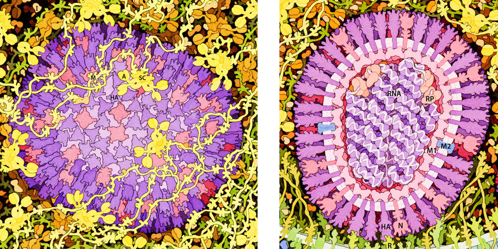
The two illustrations below highlight the overall structure of the influenza virus, with an "outside" view of the virus on the left and a cross-section "inside" view on the right. These images were created by the artist David Goodsell as part of the Flu Fight Poster.
Take some time to explore each image and try to identify the key parts of the influenza virus, including the viral membrane (sometimes called the "envelope") that surrounds the RNA genome inside. The outer surface of the virus envelope is covered in proteins, specifically the hemagglutinin protein (shown in purple with abbreviation "HA") and the neuraminidase protein (colored pink with abbreviation "N").
The influenza (flu) virus cannot survive and reproduce on its own. It needs to infect a host in order to complete the virus infection cycle. In its simplest form, this process includes three steps:
1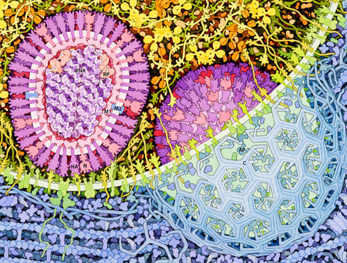 .) Attachment & Endocytosis
The virus attaches to the host cell through a process called receptor-mediated endocytosis.
Hemagglutinin proteins (shown in purple) on the outer surface of the virus attach to sialic acids (shown in green) on the surface of the host cell. Sialic acids are a common type of sugar (carbohydrate) that is found in high quantities on the surface of human epithelial cells.
The virus is then endocytosed, meaning it is surrounded by the host cell's membrane and absorbed into the host cell (shown on the right side of the image).
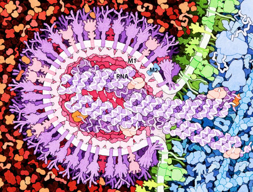
2.) Membrane Fusion & Genome Release
At this point, the entire virus has been taken into the host cell. But the viral genome is still stuck inside the viral membrane.
The same Hemagglutinin proteins that were involved in attachment now help pull the viral membrane and host cell membrane together. This results in membrane fusion and the release of the RNA genome into the host cell's cytoplasm (shown in blue)
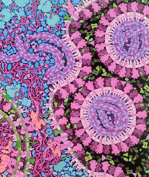
3.) Replication and Viral Budding
Once the viral genome is released into the cytoplasm of the host cell, it begins to be replicated using the host cell's own proteins.
Additional copies of key viral proteins - such as the hemagglutinin protein - are also created, being translated from the viral genome using the host cell's own proteins.
The replicated viral genomes and translated viral proteins assemble into full new influenza viruses that then bud off from the infected host cell, where they go on to infect other cells.
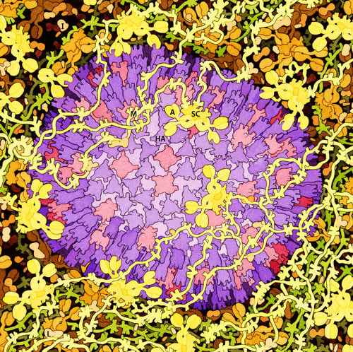 The mucosal barrier that lines the ciliated epithelial cells of the upper respiratory tract in humans is composed of a thick network of proteins that work to trap and neutralize the virus, and to activate additional antiviral responses.
Mucins (colored yellow with abbreviation "M") are long, fibrous proteins that intertwine to create a thick, viscous network.
Antibodies (colored yellow with abbreviation "A") are produced in our adaptive immune system in response to either a vaccine or a previous infection. They bind primarily to regions of the hemagglutinin protein (colored purple with abbreviation "HA") found on the surface of the influenza virus, which blocks hemagglutinin from binding to sialic acids.
Other proteins such as interferon (I) and lactoferrin (L) make up part of the innate immune system that activates other nonspeci c antiviral defense mechanisms.
When viruses aren’t neutralized by antibodies or other parts of the innate immune system, they attach to cells and begin the virus infection cycle.
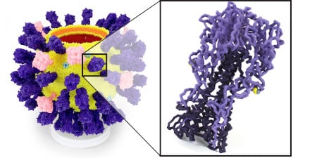 The 2026-2027 prebuild model will represent residues 153-252 of chain A of the hemagglutinin protein structure file 3UBE.pdb. This region includes the sialic acid binding domain of the hemagglutinin protein found on the surface of influenza viruses.
The same prebuild protein structure will be used at all levels of the event. Teams are encouraged to improve and refine their prebuild models as they participate in higher levels of the event, as model judging will become more rigorous.
More details on this year's prebuild protein structure, building your prebuild model, how it will be scored, and what creative additions you may want to include can be found in module 3 (https://learn.3dmoleculardesigns.com/science-olympiad-student-team-training-resources-module-3) of these training resources