
Skip to content
header-top-2
About the Event
Module 1: Protein Structure
Module 2: Jmol Training
Module 3: Prebuild Model
Onsite Exam Prep

Student Resources

Module 3: Designing the Prebuild Model


Before the event, teams will build a protein model to be impounded for judging the morning of the competition. This Prebuild Model will be worth 40% of the team's total score.

The same prebuild protein structure will be used at all levels of the event. Teams are encouraged to improve and refine their prebuild models as they participate in higher levels of the event, as model judging will become more rigorous.


What Does it Mean to "Design" a Protein Model?

Designing a physical model of a protein structure is a key component of the Science Olympiad Protein Modeling Event.

But what does it really mean to "design" a protein model? It means you explore a protein structure in a molecular visualization program, and then simplify the way the protein is visually displayed to make the key features of the protein more obvious to help communicate your molecular story.

This can mean hiding some atoms that are not important for your protein story, changing the display format of certain parts of your protein structure, or changing colors to better highlight the most important parts of the protein structures.

Annotating The Amino Acid Sequence of Your Model

  1. Delineate the Scale – Start by marking the scale on your linear model. All prebuild models for the Protein Modeling Event should represent 100 amino acids at a scale of 2cm per amino acid, for a total length of 200cm.
  2. Add Endcaps – Add a blue endcap at the beginning of your model to represent the N-terminus and a red endcap at the end of your model to represent the C-terminus.
  3. Map Secondary Structures – Mark the beginning and end of each alpha helix and beta sheet in the 100 amino acid long protein you are modeling.  These marks will guide the folding of the secondary structures.
  4. Annotate Key Sidechains – Mark the locations of any key sidechains you plan to display so that you can add them in the correct locations once you have folded your model.

Folding the 3-dimensional Structure of Your Model

  1. Fold the Secondary Structures – Using the annotations you added to your linear model, fold the two types of secondary structures.  Alpha helices are right handed spirals and beta sheets are zig-zag shapes.
  2. Fold the Tertiary Structure – Using the JUDE Jmol display as your guide, fold the overall shape of your protein structure. Rotating the digital display of your structure will allow you to view it from different angles, which will help you fine tune the folded shape of your physical model.
  3. Add Sidechains and Creative Additions – Once the overall 3-dimensional shape of your protein is folded, you can begin to add creative additions to further enhance your model.

How Your Model Will be Judged by Event Supervisors

  1. Scoring Rubrics – Event supervisors will use a prebuild scoring rubric to award points for the accuracy of your folded prebuild model. Having correctly positioned features such as the blue and red endcap, the two types of secondary structures, and the correct overall shape will earn your model points.
  2. Creative Additions – Going beyond a correctly folded protein backbone will be crucial for competing in higher levels of the competition.  Creative additions such as showing key sidechains or modeling other molecules that interact with the protein will earn your prebuild model additional points.
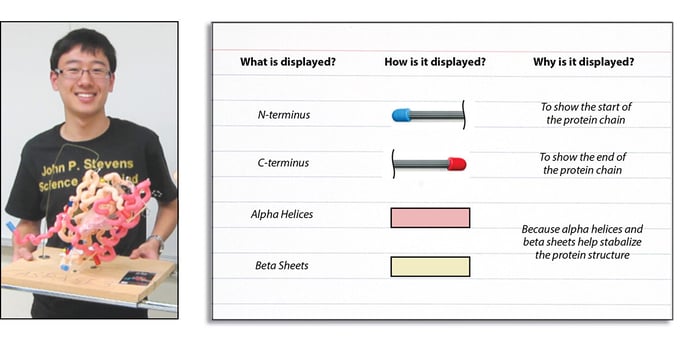
  3. The Notecard – Including a notecard that clearly describes what features you have included on your model, how you have chosen to represent those features, and why you feel they were important will earn your model points (see example below).
  4. Model Size - Prebuild models must not exceed 50cm x 50cm x 50cm
20

The 2026-2027 Prebuild Protein Structure


The 2026-2027 prebuild model should represent
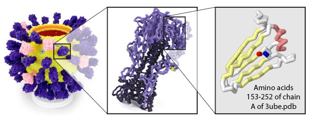
residues 153-252 of chain A of the protein structure file 3UBE.pdb 

The same prebuild protein structure will be used at all levels of the event. Teams are encouraged to improve and refine their prebuild models as they participate in higher levels of the event, as model judging will become more rigorous. 

An Overview of the 3ube.pdb Hemagglutinin Prebuild Structure

The 3ube.pdb structure file includes one complete copy of the hemagglutinin protein, the envelope protein that covers the outer surface of the influenza virus.  The hemagglutinin protein is a trimer (a protein made up of three identical monomers) that folds into a stalk-like quaternary structure.

The hemagglutinin protein is involved in both the attachment of the virus to a host cell membrane (by binding to sialic acid sugars) and the fusion of the viral membrane to the host cell membrane, allowing for the viral genome to be released. 

The specific region to be modeled, residues 153-252 of chain A of the protein structure file 3UBE.pdb, includes the sialic acid binding domain and common antigenic drift loops.
6-3


Key Features of the Prebuild Structure

11The Hemagglutinin Protein is a Trimer


The hemagglutinin protein has more than one protein chain. It is made up of three identical "monomers", each comprised of two protein chains, for a total of six chains in a full hemagglutinin protein. 


Your prebuild model should represent amino acid residues 153-252 of chain A.


7-2The Sialic Acid Sugar


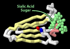
This year's prebuild represents the sialic acid binding domains in the hemagglutinin protein.  The structure file includes the sialic acid sugar itself!


8-4The Sialic Acid Binding Sidechains


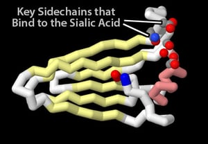
You may want to explore how the sialic acid structure fits into the hemagglutinin protein.  Specific amino acid sidechains, discussed in detail in figure 1 and figure 2 of the 3ube.pdb scientific article, interact with key regions of the sialic acid sugar to bind tightly to it.


9Antigenic Drift Regions


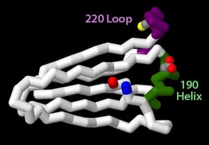
It is common to see seasonal changes (mutations) to the sialic acid binding domain of the hemagglutinin protein. Over time, these mutations allow the influenza virus to evade existing antibody recognition, overcome vaccination and sometimes even jump from infecting one species to infecting a different species (ie: swine or birds).

These key regions are discussed in detail in figure 5 of the 3ube.pdb scientific article, including the key mutations that led to the 2009 influenza virus pandemic.


10-1Trimer Cysteine Sidechains


The hemagglutinin protein has three protein chains. Proteins with more than one chain are said to have quaternary structure.


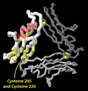
Specific cysteine amino acids in each of the three identical chains in the hemagglutinin protein are involved in holding the three chains together.  These sidechains form a specific type of covalent bond called a disulfide bond.


Prebuild Creative Additions

At all levels of the competition, the prebuild model will be judged based on two main features:

  1. The accuracy of the overall fold of the protein model’s backbone 
  2. The creative additions used to enhance the protein model 

Creative additions can make the difference between an average prebuild model and a winner.  Using modeling materials of your choice, enhance your prebuild model with features that represent key concepts and structural features related to this year's protein topic and prebuild structure. 

This year's Prebuild Model represents a fragment of the hemagglutinin protein found on the surface of influenza viruses. Some excellent area to explore for your creative additions, including:
  • The sialic acid analog in the prebuild model protein structure 3UBE.pdb
  • Important amino acid sidechains involved in binding to sialic acids
  • Important mutations to the hemagglutinin protein that allow for virus crossover and faster spread
  • Common regions of the hemagglutinin protein involved in antigenic drift
  • Cysteine amino acid sidechains involved in stabilizing the hemagglutinin protein
  • General "principles of chemistry that drive protein folding", such as a hydrophobic core, a hydrophilic shell, and salt bridges.

Note that judges can award points for other creative additions not listed here, if they are relevant to the protein structure and accurately shown.  Higher levels of the competition will award a larger portion of the prebuild model score based on creative additions.


Reading a Scientific Paper

Creative additions can also be based on information contained in the scientific paper published when this year's prebuild protein structure was discovered.  Be aware, reading a scientific paper can be challenging!  The video below provides some recommendations for how to approach this task. 

Scientific Article: https://journals.asm.org/doi/10.1128/jvi.06322-11


  1. Introduction Section:  Do not start with the abstract (usually the first section in a scientific paper). Rather, skip to the introduction section, which will provide some background for the content in the paper. 
  2. Identify Vocabulary: After reading the introduction, take some time to look up any vocabulary you are not familiar with. This will help you understand the remaining sections of the paper.
  3. Images and Image Captions: Next, explore the various figures and data diagrams throughout the paper, including the captions below each of the figures. 
  4. Conclusion Section: This section will describe the primary findings of the research that was described in more detail with the figures and captions found throughout the rest of the paper.
  5. Abstract Section: Finally, return to the abstract section, usually found at the very beginning of the paper for a wholistic overview of the research done.

About the Event
Module 1: Protein Structure
Module 2: Jmol Training
Module 3: Prebuild Model
Onsite Exam Prep