


Protein Builders Event (Division B)
Student Training Resources
The Onsite Model
During the event session, teams will build a physical model representing a 20 amino acid long protein sequence. Materials for the onsite modeling activity will be provided by the event supervisor, including:
- A 1.0 meter long Modeling Tie that will serve as the protein backbone
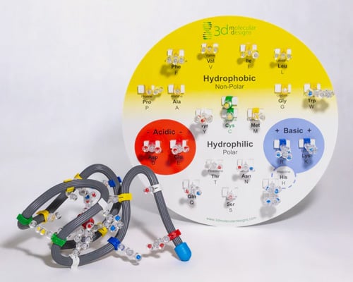
- A set of 20x Amino acid sidechain models with property-color-coding clips to attach to the Modeling Tie
- An Amino Acid Sidechain Chart that identifies each amino acid and its chemical properties
- The specific sequence of 20 amino acids to be modeled at the event
Background information on protein structure and folding can be found below. A collection of practice sequences similar to the sequences used during the competition can be downloaded here. Teams can use 3D Molecular Designs’ Amino Acid Starter Kit to practice folding protein models and experience how the onsite modeling activity will work.
The Onsite Exam

During the event session, teams will also complete a multiple-choice and short-answer written exam. The exam will include questions related to general protein structure and folding and will be based on the content on this web page and the resources linked on it.
Questions become more challenging at State and National tournaments. Use the Study Guide by Tournament Level near the end of this page to see what to focus on.
Each participant is allowed to bring one 8.5 x 11 sheet of paper, which may be in a sheet protector sealed with tape or laminated, and may contain information on both sides in any form and from any source, without any affixed labels. Teams should bring a ruler or meter stick to help properly space the amino acid sidechains on their Modeling Tie.
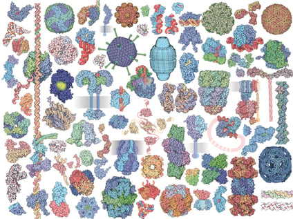
Proteins are some of the most important molecules in our body. They are made using nutrients we take in from our food and perform a countless variety of tasks in our body. A few examples include:
-
structural components in hair or nails (keratin proteins) and skin (collagen proteins)
-
contracting muscles (actin and myosin proteins)
-
sending brain signals (channel proteins and neurotransmitter receptor proteins)
- carrying oxygen in our blood (hemoglobin proteins)
-
replicating cells (polymerase proteins)
There are a wide variety of different types of protein molecules. Each type of protein has a different shape or structure that allows it to perform a different task or function.
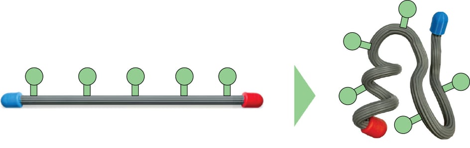
Protein molecules are made of smaller molecular units called amino acids. These individual amino acids are joined together to form a long protein chain, just like individual links of a metal form a long strand of flexible chain.

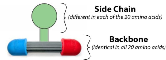
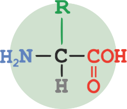
Each protein chain has a beginning, called the N-terminus (colored blue in the illustration above) and an end, called the C-terminus (red). This gives the protein chain directionality, with a beginning and an end. Each amino acid in a protein chain has a backbone region (gray) and a sidechain (green) that sticks out from the backbone.
The linked amino acids in a protein chain do not stay a straight linear molecule. Rather, they fold and bend into complex shapes. In this way, a simple sequence of amino acids linked together can take on a very complicated 3-dimensional structure! This folded structure determines what the protein does and how it functions.
The Twenty Types of Amino Acids
There are twenty* different types of amino acids that can be linked together in a protein sequence. In each of the twenty types, the backbone is identical, but the sidechain is different.
Below is a table showing all twenty amino acids. Notice how the backbone region in each amino acid is the same but the sidechain in each amino acid is different. This is because the atoms that make up each sidechain are different, with carbon atoms shown as gray circles, nitrogen atoms shown as blue circles, oxygen atoms shown as red circles and sulfur atoms shown as yellow circles.
Take some time to study this table. Can you find any patterns? What types of atoms make up the different sidechains? What types of shapes do the different sidechains have?
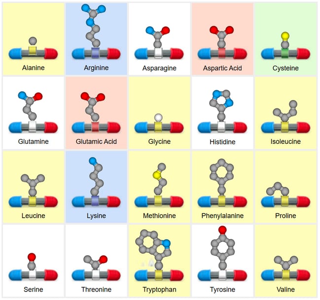
The twenty sidechains can be grouped into four main categories based on the shape and type of atoms in their sidechain. These categories impact how a protein folds into a 3-dimensional shape.
Hydrophobic (non-polar)
Primarily made up of carbon atoms, these sidechains like to hide away from water at the center of a folded protein chain.
In the on-site event, hydrophobic (non-polar) side- chains will have yellow clips.
Hydrophilic (polar)
Having some nitrogen and oxygen atoms, these side- chains like to interact with water on the outside of a folded protein chain.
In the on-site event, hydrophilic (polar) sidechains will have white clips
Positive and Negative
Having lots of nitrogen atoms (positive) or lots of oxygen atoms (negative), these side- chains like to interact with each other in a folded protein chain.
In the on-site event, positive sidechains will have blue clips and negative sidechains will have red clips.
Cysteine
Cysteine is a unique amino acid with a single sulfur atom. Cysteine sidechains like to interact with each other in a folded protein chain.
In the on-site event, cysteine sidechains will have green clips.
*Most proteins are built from 20 standard amino acids; a rare 21st, selenocysteine, is found in a few specialized proteins, but for simplicity we focus on the 20 standard amino acids.
Two Sidechains with Special Shapes: Glycine and Proline
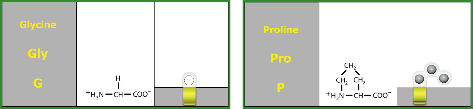
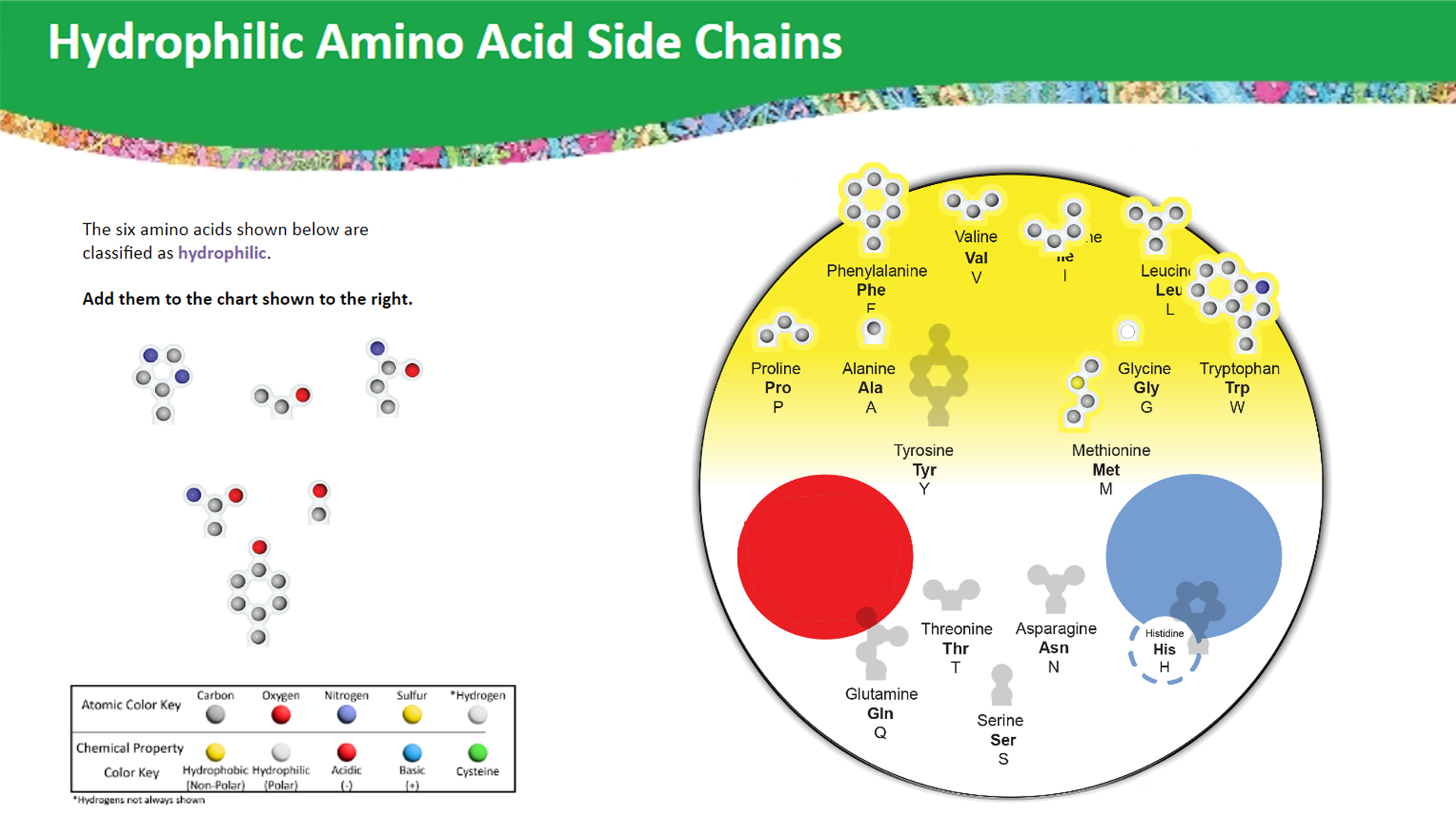
Glycine and proline have yellow (hydrophobic) clips, but their shapes give them special jobs.
Glycine has the smallest sidechain: a single hydrogen atom. This lets the chain bend and twist easily, so glycine is often found in tight turns.
Proline’s sidechain loops back and bonds to its own backbone, forming a ring. This makes the chain stiff and puts a kink in it, so proline often breaks up alpha helices.
In order to successfully build your on-site protein model, you will need to understand the four categories of sidechains (hydrophobic (non-polar), hydrophilic (polar), positive/negative and cysteine) and how each category impacts protein folding.
At the beginning of each on-site Protein Builders event, teams will be given a specific 20x amino acid long sequence to model. Teams will need to add the correct amino acid sidechains to their backbone Modeling Tie wire and then fold the wire into a 3-dimensional shape that properly follows the four principles of protein folding.
The sequence of amino acids in a protein determines what shape it folds into.
Try these free online activities. Sort the amino acids, then find them in real proteins you can rotate in 3D.
Side Chain Properties Interactable
Sort all 20 amino acids and find the core, surface, salt bridges, and disulfide bond in a real protein.
Practice Now
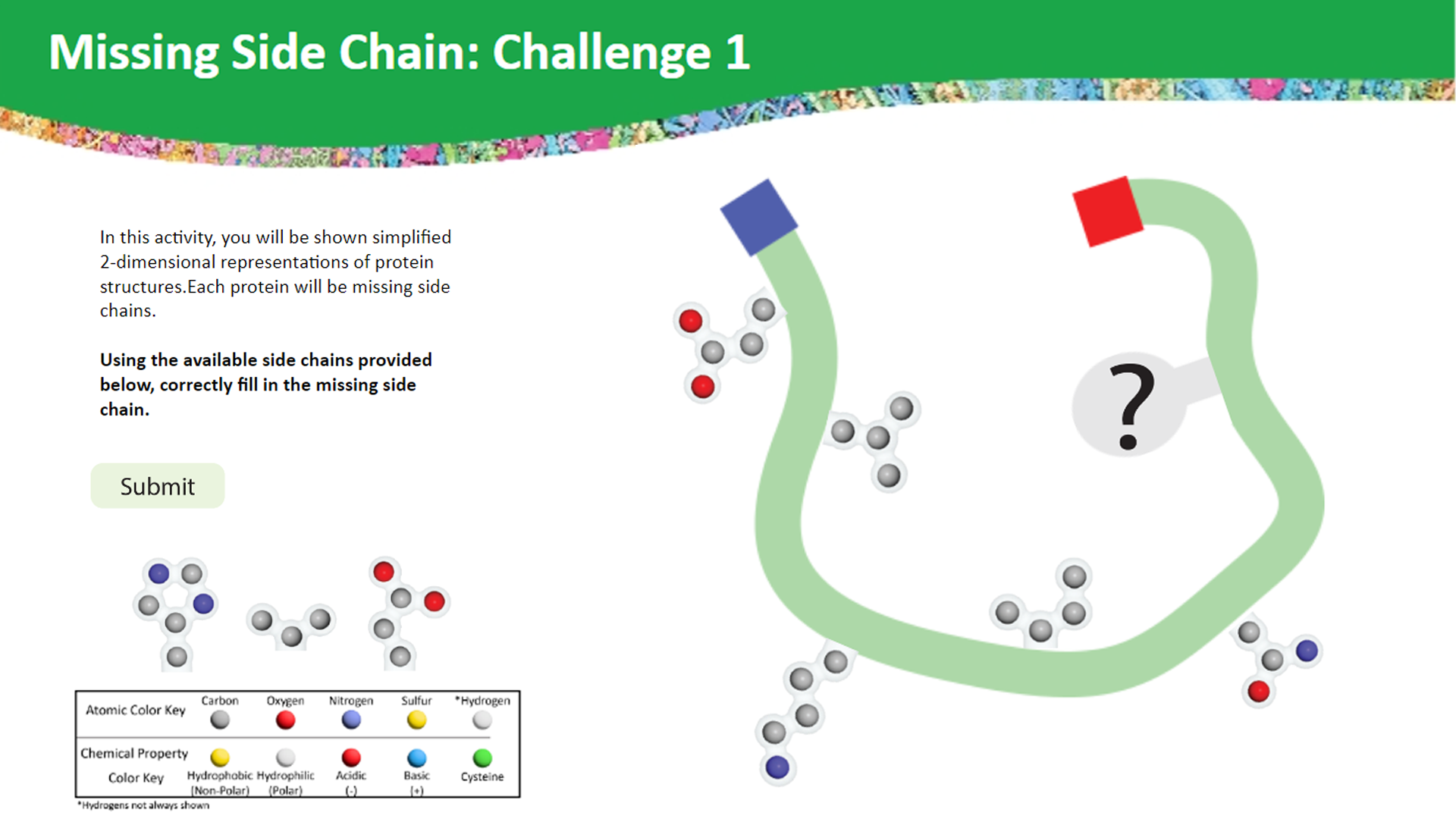
Side Chain & Protein Property Interactable
Five challenges: fill in the missing sidechain in five real proteins.
Practice Now
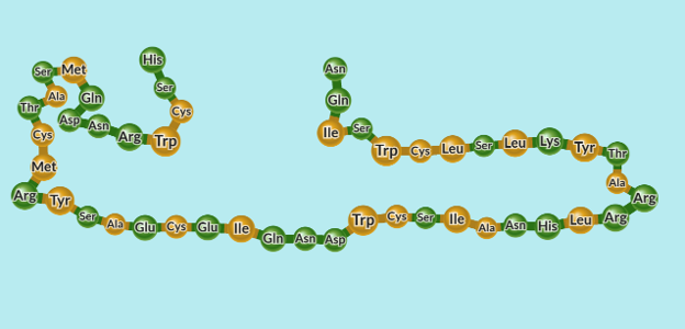
The Four Levels of Protein Structure
Primary Structure
The sequence of amino acids, from the N-terminus to the C-terminus.
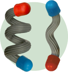
Secondary Structure
Parts of the chain fold into alpha helices and beta sheets.
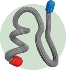
Tertiary Structure
Helices, sheets, and loops pack into one overall 3D shape.
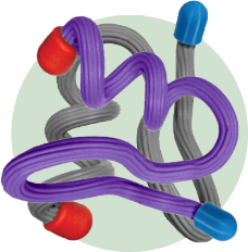
Quaternary Structure National
Some proteins are made of more than one chain. The chains fit together using the same four principles.
Helices and Sheets

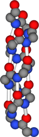
An alpha helix is a right-handed spiral. Hydrogen bonds between backbone atoms, about four amino acids apart, hold each turn in place.
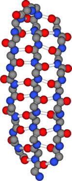
A beta sheet forms when stretches of the chain line up side by side. Hydrogen bonds between neighboring strands hold the sheet together.
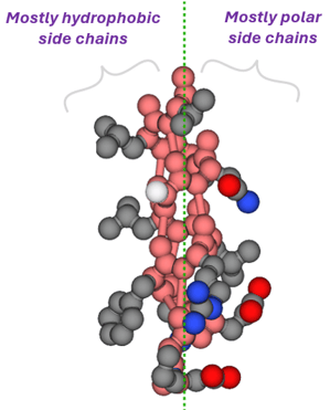
Look at where the sidechains point. Many helices and sheets have hydrophobic sidechains on one side and hydrophilic sidechains on the other. Scientists call these amphipathic. The hydrophobic side faces the protein’s core, and the hydrophilic side faces the water.
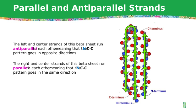
Strands that run in opposite directions are antiparallel.
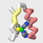
Practice folding an alpha helix and a beta sheet to model a Zinc Finger protein motif.
The Forces That Hold a Protein Together
Hydrogen bonds: A hydrogen atom on a nitrogen or oxygen is attracted to another nearby nitrogen or oxygen. Each one is weak, but proteins have many. They hold helices and sheets together, and hydrophilic sidechains such as serine and asparagine can form them with water or other sidechains.
Hydrophobic effect: Hydrophobic sidechains cluster together, away from water.
Salt bridges: A positive sidechain and a negative sidechain attract each other. This is an ionic interaction.
Disulfide bonds: Two cysteine sidechains form a covalent bond. This is the strongest of the four.
Van der Waals attractions (National): Very weak attractions between atoms that are very close together. They help tightly packed sidechains in the core stick together. Larger sidechains have more of them.
ACS Middle School Chemistry: Water Is a Polar Molecule
Background on polarity.
Concord Consortium: Hydrogen Bonds State
Resource for preparing for the State competition.
Concord Consortium: Comparing Dipole-Dipole to London Dispersion National
Weak attractions between molecules.
Concord Consortium: Factors Affecting London Dispersion Attractions National
How size and shape change van der Waals attractions.
Where a Protein Folds: Water or Membrane
State & National
Most proteins fold in water. Their hydrophobic sidechains form a core in the center, and their hydrophilic and charged sidechains cover the surface.
Some proteins cross a cell membrane. The middle of a membrane is oily (nonpolar). The part of the protein inside the membrane has hydrophobic sidechains on its outer surface, where they touch the membrane.
Side Chain & Protein Property Interactable — Challenge 4: Aquaporin
Membrane protein challenge.
Concord Consortium: Protein Folding
Switch the solvent from water to oil and watch how the chain folds.
Ask A Biologist: Protein Channels
Channel proteins in the cell membrane.
When One Amino Acid Changes
All levels (required by the rules)
A change in the DNA can swap one amino acid for another. Even one change can affect how a protein folds and works. To predict the effect, ask: What category was the old sidechain? What is the new one? Where is it in the folded protein?
Try it with Practice Sequence 1:
- Leu (position 4) → Asp: A charged sidechain is now in a spot that should be in the hydrophobic core. The protein may not fold correctly.
- Cys (position 10) → Ser: The disulfide bond with Cys 16 can no longer form. The protein is less stable.
- Glu → Val in hemoglobin (sickle cell): A charged surface sidechain becomes hydrophobic. Hemoglobin molecules stick together and form stiff fibers.
Some changes have little effect, for example when a sidechain swaps for another in the same category on the surface. Others are harmful, and a few are helpful.
Amino Acid Starter Kit Additional Activities (see "The Effects of Mutations")
Hands-on mutation activity using the same Amino Acid Starter Kit.
Learn.Genetics: Mutate a Gene
Make your own mutations and see how they change the protein.
Amoeba Sisters: Mutations (Updated)
Short video on the types of mutations and their effects.
ß-Globin Folding Kit Student Overview
How one amino acid change causes sickle cell disease.
PDB-101 Molecule of the Month: Hemoglobin State, National
Hemoglobin's structure and how the sickle cell change affects it.
Concord Consortium: Mutations National
Explore how different mutations change a protein.
When Conditions Change: Heat and pH
State & National
Heat makes atoms move faster. Too much heat can shake apart the weak interactions that hold a protein’s shape. The protein unfolds (denatures) and stops working. Some proteins refold when conditions return to normal.
pH measures how acidic or basic a solution is. Acidic and basic sidechains carry charges. When the pH changes, some of these sidechains gain or lose their charge, so salt bridges can break.
Ask A Biologist: Breaking Proteins
Egg-white activity: see what heat, alcohol, and lemon juice do to proteins.
Amino Acid Starter Kit Additional Activities (see "Reversible Denaturation")
Hands-on denaturation activity using the Amino Acid Starter Kit.
Amoeba Sisters: Enzymes (Updated)
Short video on how temperature and pH affect enzymes.
ACS Middle School Chemistry: pH and Color Change
Background on pH: what acidic and basic mean.
Shape and Function: Binding Sites and Active Sites
State & National
Many proteins work by binding another molecule. A binding site is a pocket shaped by sidechains. In an enzyme, this pocket is called the active site, where a chemical reaction is sped up.
Binding follows the same four principles. Hydrophobic parts of a molecule fit against hydrophobic sidechains, and charged parts form salt bridges. Changing one sidechain in a binding site can change what binds.
Binding Shape Interactable (basic binding) State
Interactive: see how shape decides what fits in a binding site.
Binding Chemical Interactable State, National
Interactive: see how hydrophobic and charged sidechains decide what binds.
Active Sites with a Mini Toober (handout) National
Build an active site from serine, histidine, and glutamic acid.
From DNA to Protein: Reading a Codon Chart
National
DNA stores the instructions for each protein’s amino acid sequence. The DNA code is copied into messenger RNA (mRNA). Each group of three mRNA bases, called a codon, codes for one amino acid. A codon chart shows which amino acid each codon codes for.
At National tournaments, you may use a codon chart to translate a short DNA sequence, or to figure out how a DNA change alters the amino acid sequence. For example, in sickle cell, the codon GAG becomes GTG, and glutamic acid becomes valine. A codon chart will be provided.
Decoding DNA with Codon Charts National
Worksheet: translate DNA with a codon chart, including the sickle cell mutation.
Decoding Beta-Globin mRNA with Codon Charts National
Worksheet: an easier starting point for reading a codon chart.
Amoeba Sisters: How to Read a Codon Chart National
Video: step-by-step on using a codon chart.
Amoeba Sisters: Codon Chart Video Companion National
Printable practice to use with the codon chart video.
Learn.Genetics: Transcribe and Translate a Gene National
Interactive with videos: transcribe DNA into mRNA and translate it into protein.
Concord Consortium: DNA to Protein National
Interactive: follow a gene from DNA to mRNA to protein.
Study Guide by Tournament Level
All levels
Exam questions build from Regional to State to National. Each level includes everything from the levels before it.
| Topic | Regional & Invitational | State | National |
|---|---|---|---|
| Levels of structure | Primary, secondary, and tertiary structure | How the sequence determines the fold | Connect all levels; quaternary structure |
| Sidechain properties | Sort sidechains with the chart; glycine, proline, and cysteine | Predict core vs. surface | Size, ring-shaped sidechains, amphipathic patterns |
| Interactions | Name the four: hydrophobic effect, hydrogen bonds, salt bridges, disulfide bonds | Explain which interaction holds a pair or region | How several work together; van der Waals |
| Helices and sheets | Recognize helices, sheets, and loops | Backbone hydrogen bonds; glycine and proline effects | Parallel vs. antiparallel; amphipathic helices |
| Where proteins fold | Soluble protein in water | Core vs. surface; membrane segment | Compare water and membrane |
| Substitutions | One change with an obvious effect | Compare two changes | Several changes or two versions |
| Heat and pH | Water vs. oily environments | pH can break salt bridges | pH, heat, and unfolding together |
| Diagrams | Simple helix and sheet drawings | Label termini and find residues | Ribbon and space-filling images |
| Function | Shape determines function | Binding sites and active sites | Binding tied to specific sidechains |
| DNA to protein | — | — | Use a codon chart; trace a DNA change |
Topics you do not need to study:
- Molecular visualization software (JUDE or Jmol) and PDB files
- The Protein Modeling (Division C) theme protein
- Changes made to proteins after they are built (post-translational modifications)
- How scientists determine protein structures (X-ray, cryo-EM, NMR)
- Enzyme kinetics, lock-and-key and induced-fit models, and allostery
- pKa, Henderson–Hasselbalch, net charge, or isoelectric point calculations
- Memorizing sidechain structures or the codon table (both are provided)
A final reminder: When you answer an exam question, ask yourself: What do I observe in the structure? What chemical properties or interactions are involved? How could those features support the protein’s function?
Additional Protein Structure Resources
Amino Acid Starter Kit
Learn about protein structure and function with the hands-on kit that inspired the Protein Builders event.
What is a Protein?
Visualize what proteins are, how they fold, and how their 3-dimensional structure relates to their function.
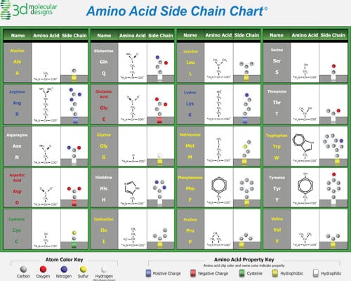
Amino Acid Side Chain Chart
Explore the structures of each of the twenty amino acids using this detailed amino acid side chain chart.
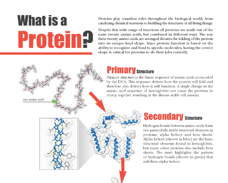
What Is a Protein? Flyer
A one-page guide to protein structure from PDB-101, including how one amino acid change causes sickle cell anemia. Image: RCSB PDB (PDB-101), CC BY 4.0.
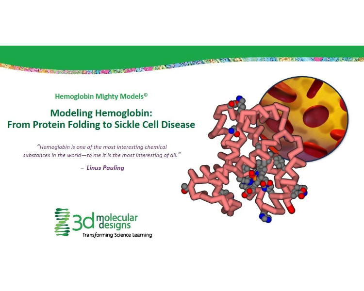
Digital Modeling Hub
Free 3D Molecular Designs interactables, slide decks, and handouts for exploring proteins.
Additional Links and Resources
- Practice Kits: Amino Acid Starter Kit©
- Division B Official Rules: 2027 Rules | Science Olympiad
